


Our aim was two-fold, to study the interobserver agreement in tumour segmentation and to search for a reliable methodology to segment gliomas using 18F-Fluorocholine PET/CT.
Methods25 patients with glioma, from a prospective and non-randomized study [FuMeGA (Functional and Metabolic Glioma Analysis)], were included.Interobserver variability in tumour segmentation was assessed using fixed thresholds. Different strategies were used to segment the tumours. First, a semi-automatic tumour segmentation was performed, selecting the best SUVmax-% threshold for each lesion. Next we determined a variable SUVmax-% depending on the SUVmax. Finally a segmentation using a fixed SUVmax threshold was performed. To do so, a sampling of 10 regions of interest (ROIs of 2.8cm2) located in the normal brain was performed. The upper value of the sample mean SUVmax±3 SD was used as cut-off. All procedures were tested and classified as effective or not for tumour segmentation by two observer's consensus.
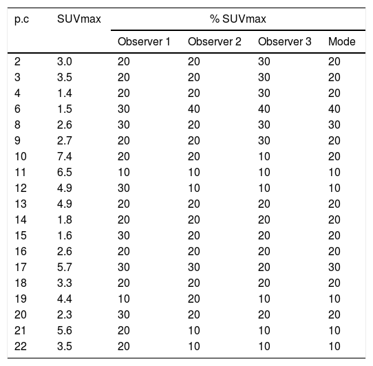
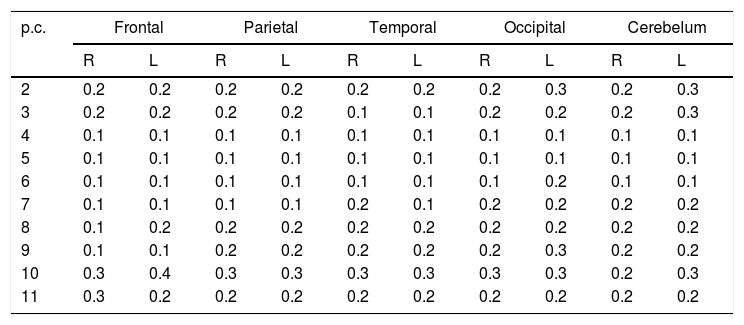
ResultsIn the pilot segmentation, the mean±SD of SUVmax, SUVmean and optimal SUVmax-% threshold were: 3.64±1.77, 1.32±0.57 and 21.32±8.39, respectively. Optimal SUVmax-% threshold showed a significant association with the SUVmax (Pearson=−0.653, p=0.002). However, the linear regression model for the total sample was not good, that supported the division in two homogeneous groups, defining two formulas for predicting the optimal SUVmax-% threshold. As to the third procedure, the obtained value for the mean SUVmax background+3 SD was 0.33. This value allowed segmenting correctly a significant fraction of tumours, although not all.
ConclusionA great interobserver variability in the tumour segmentation was found. None of the methods was able to segment correctly all the gliomas, probably explained by the wide tumour heterogeneity on 18F-Fluorocholine PET/CT.
El objetivo fue doble, valorar el acuerdo interobservador en la segmentación tumoral y la búsqueda de una metodología fiable y aplicable en la segmentación de gliomas usando PET/TC con 18F-Fluorocolina.
Material y métodosSe incluyeron 25 pacientes con glioma, procedentes de un estudio prospectivo no randomizado [FuMeGA (Functional and Metabolic Glioma Analysis)].Se analizó la variabilidad interobservador usando umbrales fijos. Diferentes estrategias se emplearon en la segmentación. Primero, se realizó una segmentación semiautomática, seleccionando el mejor umbral del % de SUVmax para cada lesión. Posteriormente, se determinó una variable del % de SUVmax dependiente del SUVmax. Finalmente se realizó una segmentación usando un valor de umbral fijo de SUVmax. Para ello, se realizó un muestreo de 10 regiones de interés (ROIs de 2,8cm2) localizadas en cerebro normal. El valor superior obtenido de la media del muestreo±3 desviaciones estándar se usó como valor de corte. Todos los procedimientos fueron testados y clasificados como válidos o no en la segmentación tumoral en consenso por dos observadores.
ResultadosEn la segmentación piloto, la media±DE del SUVmax, SUVmedio y el umbral del %SUVmax fue de 3,64±1,77; 1,32±0,57 y 2,132±8,39, respectivamente. El valor óptimo del umbral %SUVmax mostró una asociación significativa con el SUVmax (Pearson=-0,653; p=0,002). Sin embargo, el modelo de regresión lineal del total de la muestra no fue bueno lo que justificó la división de la misma en dos grupos homogéneos, definiendo dos formulas para predecir el umbral del %SUVmax. Para el tercer procedimiento, el valor obtenido de la media SUVmax+3 DE fue de 0,33. Este valor permitió segmentar correctamente una elevada proporción de casos, aunque no todos.
ConclusiónSe encontró una gran variabilidad interobservador en la segmentacion tumoral. Ninguno de los métodos fue capaz de segmentar correctamente todos los gliomas probablemente debido a la amplia heterogeneidad en la PET/TC con 18F-Fluorocolina.
Article
If you experience access problems, you can contact the SEMNIM Technical Secretariat by email at secretaria.tecnica@semnim.es or by phone at +34 619 594 780.

Revista Española de Medicina Nuclear e Imagen Molecular (English Edition)
