


The use of contrast-enhanced spectral mammography (CESM) has increased in recent years, as has awareness of radiation dose safety among professionals and patients. The principal aim of this study was to compare radiation exposure measured using the entrance surface dose (ESD) and the average glandular dose (AGD) in CESM, Full-Field Digital Mammography (FFDM) and Digital Breast Tomosynthesis (DBT). Our second objective was to evaluate differences caused by compressed breast thickness, compression force and patient age.
MethodsA retrospective observational study included all patients who had undergone a CESM between May 2021 and May 2022. Data was collected on ESD and AGD from the different CESM studies, and breast density and volume were determined by two expert radiologists. The comparative analysis focused on the dose of radiation received during the craniocaudal (CC) projection of the right breast in CESM studies and FFDM or DBT, performed within a 12-month period. Lastly, a statistical analysis was performed to determine the influence of breast compression thickness, compression force and patient age.
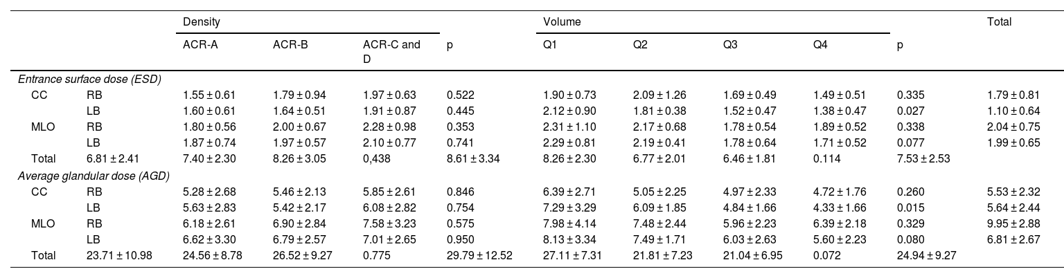
ResultsSeventy-seven patients participated in the comparative study and forty-four in the dosimetric study. Differences in radiation dose (ESD/AGD) were found among the three breast imaging techniques. The dose in CESM (1.70/5.39 mGy) was lower than in DBT (2.19/6.79 mGy) and higher than in FFDM (1.26/4.06 mGy) for an average breast compression thickness of 59.64 mm. A positive correlation was observed between the dose received in CESM and breast compression thickness (ρ = 0.55), and a negative correlation was observed with patient age (ρ = −0.27). No differences in dosimetric variables were observed for different compression forces.
ConclusionsThe ESD and AGD in the CC projection of the right breast in CESM are higher than in FFDM but lower than in DBT. The dose had a positive correlation with breast compression thickness and a negative correlation with patient age.
El uso de la mamografía espectral con contraste (CEM) ha aumentado en los últimos años, así como la concienciación sobre la dosis de radiación por parte de los profesionales y pacientes. El objetivo principal del estudio es comparar la dosis de radiación, medida como dosis de entrada (DE) y dosis media glandular (DMG) de la CEM, mamografía digital y tomosíntesis mamaria. El objetivo secundario fue evaluar las diferencias según el espesor de la mama en compresión, la fuerza de compresión y la edad de la paciente.
Material y métodosEstudio retrospectivo observacional en el que se incluyeron las pacientes a las que se les había realizado una CEM entre mayo del 2021 y mayo del 2022. Se recogieron la DE y la DMG de las distintas proyecciones de la CEM y se determinó la densidad y volumen mamarios por dos radiólogos expertos. Para el análisis comparativo, se valoró la radiación recibida en la proyección craneocaudal (CC) de la mama derecha de los estudios de CEM y de mamografía convencional o tomosíntesis mamaria realizados en un intervalo de tiempo menor de un año. Por último, se realizó un análisis estadístico para determinar la influencia del espesor de la mama en compresión, la fuerza de compresión y la edad de la paciente.
ResultadosSetenta y siete pacientes participaron en el estudio comparativo y cuarenta y cuatro en el estudio dosimétrico. Se encontraron diferencias en la dosis de radiación (DE/DMG) entre las tres técnicas de imagen mamaria. En CEM (1,70/5,39 mGy) fue menor que en tomosíntesis (2,19/6,79 mGy) y mayor que en mamografía convencional (1,26/4,06 mGy) para un espesor medio de mama en compresión de 59,64 mm. Se obtuvo una correlación positiva entre la dosis recibida en la CEM y el espesor de la mama en compresión (ρ = 0,55), y una negativa con la edad de la paciente (ρ = −0,27). No se apreciaron diferencias en las variables dosimétricas para distintas fuerzas de compresión.
ConclusionesLa DE y DMG en la proyección CC de la mama derecha en CEM es mayor que en mamografía, pero menor que en tomosíntesis. La dosis aumentó con el espesor de la mama en compresión y disminuyó con la edad de la paciente.

