


Since mechanical thrombectomy has allowed ischaemic stroke thrombus retrieval, the exhaustive study of this material has enabled better understanding of the potential physiopathological processes involved in thrombus formation.
DevelopmentThrombotic pathways involved in the different vascular beds share common mechanisms, causing difficulties in the identification of specific patterns associated with stroke aetiology. However, other factors such as clot formation time, associated inflammatory status, or activation of additional immune and coagulation pathways (neutrophil extracellular trap [NET] delivery, platelet aggregation, endothelial activation, and von Willebrand Factor release) have been described as determinants in thrombus characteristics. Thus, variable proportions of fibrin-/platelet-rich and erythrocyte-rich areas are closely interrelated within the thrombus, frequently associated with a protective outer shell with high concentrations of fibrin, NETs, and von Willebrand Factor. The presence of these components, as well as their distribution and interrelationships, have been shown to have effects on the thrombus’ resistance to revascularisation treatments. Understanding of these pathways has enabled the development of adjuvant therapies capable of enhancing current fibrinolytic drugs and/or increasing the efficacy of endovascular treatments.
ConclusionUnderstanding of thrombus components and mechanisms involved in thrombus formation represent a potential pathway for the development of ischaemic stroke therapeutics with promising perspectives.
La obtención del material trombótico en el ictus isquémico desde la generalización de la trombectomía mecánica ha permitido un estudio exhaustivo del mismo y de los posibles mecanismos fisiopatológicos subyacentes a su formación.
DesarrolloLos mecanismos trombóticos implicados en los diferentes lechos vasculares no difieren en exceso, condicionando una heterogeneidad importante que ha dificultado la identificación de patrones asociados al origen o etiología del ictus. Se ha observado sin embargo una mayor influencia de factores como el tiempo de desarrollo del coágulo, el estado inflamatorio sistémico o la activación de vías inmunes y de la coagulación [liberación de Neutrophil extracellular traps (NETs), agregación plaquetar, activación endotelial y liberación de Factor de vonWillebrand]. Como resultado, en el seno del trombo conviven en proporciones variables áreas ricas en eritrocitos y áreas ricas en plaquetas/fibrina con una íntima relación entre ellas y acompañadas con frecuencia de una coraza protectora con alto contenido en fibrina, Factor vonWillebrand y NETs. La presencia de estos componentes, así como su disposición e interrelación ha demostrado tener efectos en la resistencia a los diferentes tratamientos revascularizadores. El conocimiento de estas vías ha permitido el desarrollo de posibles terapias adyuvantes con capacidad de potenciar los tratamientos fibrinolíticos actuales y/o aumentar la eficacia del tratamiento endovascular.
ConclusiónEl conocimiento de los componentes del trombo y los posibles mecanismos implicados en su formación es una potencial vía de desarrollo en el tratamiento del ictus isquémico con resultados prometedores.
Stroke is one of the main causes of disability and death in our setting,1 with ischaemic stroke, which is caused by an obstruction to arterial flow in brain structures, representing 80% of strokes.2 The main occlusive agent in cerebral arteries is usually thrombotic material derived from thromboses of several aetiologies. Extraction of thrombi by endovascular procedures3 has enabled detailed analysis of thrombotic material and an exponential growth of studies on the histopathological analysis of thrombi. Consensus documents have also been published to standardise the processes for obtaining, manipulating, and studying thrombi.4
This document reviews the current state of the research into thrombi in ischaemic stroke, as well as some of its applications: understanding of pathophysiological processes underlying their formation; the identification of patterns associated with strokes of different aetiologies enabling their identification in situations of uncertainty (undetermined stroke); the analysis of predictors of response to current revascularisation treatments; and new potential therapies to improve or replace current treatments.
DevelopmentThrombus composition and underlying pathophysiological processA series of agents are involved in the thrombotic process that interact in different degrees in each case, until the eventual formation of an occlusive thrombus. The mechanisms participating in the thrombosis/fibrinolysis imbalance that lead to the formation of the clot are varied, which explains the great heterogeneity observed in histopathological analysis of thrombi.6
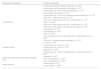
One of the most widely studied factors is the location and the conditions in which the thrombus is formed and how the physical/chemical environment in which it is formed impacts its composition. In thrombi formed in the arterial bed, secondary to endothelial damage, platelet activation by tissue factor5 and the action of the von Willebrand factor (VWF)6 lead to an early involvement of thrombin and rapid formation of an extensive fibrin mesh whose cross-linking is mediated by platelet factor XIII (FXIII), which is released after activation.7 This phenomenon occurs in situations of rapid arterial blood flow that favour the formation of layers with an intricate interface between platelet- and fibrin-rich areas, known as the lines of Zahn (Fig. 1).8 However, this mechanism is not unique, and blood stasis related to atheromatous plaques or anatomical alterations,9 as well as the prothrombotic environment associated with the typical inflammatory status of systemic atheromatosis, are also present.10 In the heart cavities, the participating mechanisms include the activation of the extrinsic pathway due to damage to the endocardium, a context of blood stasis, and the loss of haemostatic balance in favour of a prothrombotic situation.11 The participating mechanisms in both areas are therefore similar but also varied, which supports part of the heterogeneity and the overlap observed in the studies performed.12
, Martius Scarlet Blue (MSB), specific immunostaining for von Willebrand factor (VWF), and specific immunostaining for Cd42b+ platelets. Platelet-rich areas are marked with asterisks (*) and erythrocyte-rich areas with a hash sign (#). The marked areas are shown at 20× magnification in the lower right corner of each image. Source: Atherothrombosis Laboratory, CIMA/Universidad de Navarra.")
Histological sections of an ischaemic stroke thrombus stained with: haematoxylin and eosin (HE), Martius Scarlet Blue (MSB), specific immunostaining for von Willebrand factor (VWF), and specific immunostaining for Cd42b+ platelets. Platelet-rich areas are marked with asterisks (*) and erythrocyte-rich areas with a hash sign (#). The marked areas are shown at 20× magnification in the lower right corner of each image.
Source: Atherothrombosis Laboratory, CIMA/Universidad de Navarra.
Many authors have sought to associate the relative content of each of the main thrombus components with the aetiology of stroke, obtaining diverging results (Table 1). Larger series published in recent years13–15 suggest increased presence of erythrocytes in atherosclerotic thrombi, and especially in those secondary to processes of vascular dissection or atheromatous plaque rupture. They also point to a higher concentration of fibrin and platelets in thrombi of cardiac origin, though without ruling out the possibility that the latter situation may be due to previous anticoagulation treatment.16 Nevertheless, the great variability observed seems to prevent aetiological classification considering only those parameters. In this regard, based on the largest series published to date, Brinjikji et al.15 acknowledged that they had not identified clinically significant differences distinguishing between cardioembolic and atherothrombotic aetiology classified according to typical histological staining, despite finding statistically significant differences, given the large sample size (1350 thrombi).
Thrombus composition and stroke aetiology.
| Aetiological classification | Thrombus composition |
|---|---|
| Cardioembolic | ↔ erythrocytes and fibrin between aetiologies, N = 2591 |
| ↔ erythrocytes and fibrin between aetiologies, N = 5092 | |
| ↑ erythrocytes with ↔ fibrin between aetiologies, N = 1793 | |
| ↔ fibrin between aetiologies, N = 2232 | |
| ↑ erythrocytes and ↓ fibrin with ↔ platelets between aetiologies, N = 3794 | |
| ↑ fibrin and ↑ inflammatory cells, N = 3228 | |
| ↑ fibrin and ↑ inflammatory cells with ↓ erythrocytes, N = 13713 | |
| ↑ NETs, N = 6826 | |
| ↑ fibrin and ↑ inflammatory cells with ↓ erythrocytes, N = 18770 | |
| ↑ fibrin and ↔ inflammatory cells between aetiologies, N = 7964 | |
| ↑ erythrocytes, N = 3766 | |
| ↑ macrophages, N = 8595 | |
| ↑ NETs, N = 8033 | |
| ↑ fibrin and platelets and ↓ erythrocytes with ↔ inflammatory cells, N = 135015 | |
| Atherothrombotic | ↑ fibrin with ↔ platelets between aetiologies, N = 3794 |
| ↑ T cells, N = 5434 | |
| ↑ erythrocytes and ↓ fibrin, N = 3228 | |
| ↑ erythrocytes and ↓ fibrin, N = 7964 | |
| ↑ platelets, N = 10596 | |
| ↑ erythrocytes and ↓ fibrin and platelets with ↔ inflammatory cells, N = 135015 | |
| Stroke associated with cancer/hypercoagulable state | ↑ platelets, N = 3249 |
| ↑ fibrin and platelets, N = 15250 | |
| ↑ fibrin and platelets, N = 135015 | |
| Artery dissection | ↑ erythrocytes, N = 2232 |
| ↑ erythrocytes, N = 135015 |
NETs: neutrophil extracellular traps.
In addition to the predominant components of a thrombus (erythrocytes, platelets, and fibrin), the thrombus matrix includes other significant elements in ischaemic stroke. One of the most widely studied elements is VWF, a glycoprotein involved in the activation and adhesion of platelets with one another and with the subendothelial matrix. Its presence has been associated with platelet, leukocyte, and fibrin concentration,17,18 and is inversely correlated with erythrocyte concentration,19 distributed particularly in platelet-rich areas and at their borders close to fibrin-rich areas (Figs. 1 and 2).20 This relationship of fibrin and VWF in platelet-rich regions suggests a direct interaction between these 2 molecules, with a potential to stabilise the thrombus, which may influence the fibrinolytic resistance observed in some thrombi. Additionally, VWF is involved in the leukocyte recruitment process in the thrombus and leukocyte adhesion to the remaining integrin-mediated structures.21

In recent years, the concept of immunothrombosis has focused attention on the effects of inflammatory cells and molecules on the processes of thrombosis and clot stabilisation. Thus, the main inflammatory cells, leukocytes, have shown to promote mechanisms of both venous and arterial thrombosis, through the exposure to procoagulant factors, release of cytokines with platelet activation capacity, and especially through the release of accumulations of chromatins, histones, and DNA in the form of neutrophil extracellular traps (NETs).22 Leukocytes in the thrombus are located in the interface between platelet-rich and erythrocyte-rich areas (Figs. 2 and 3),18,20 and higher concentrations have been associated with cardioembolic aetiology in some studies (Table 1); however, we cannot rule out the influence of the age of the thrombus in this association. Secondary platelet activation and recruitment of new inflammatory cells by NETs have shown to increase thrombus complexity and resistance to tissue plasminogen activator (tPA),23 and to participate in the formation of the external layer of the thrombus with anti-fibrinolytic capacity.24 Furthermore, colocalisation of NETs and VWF has been observed in the core of stroke thrombi,20 suggesting an interaction between both molecules with a proinflammatory and prothrombotic effect. The presence of NETs seems to decrease the activity of ADAMTS13, a metalloprotease involved in the fragmentation and regulation of VWF, favouring the accumulation of VWF and its activation.25 Likewise, other markers of neutrophil activity, such as neutrophil elastase26 or calprotectin, are reported to be present in ischemic stroke thrombi, with calprotectin (S100A9) associated with platelet, leukocyte or neutrophil eslastase concentrations and distribution (Fig. 3).27
, CD3+ lymphocytes, CD45+ leukocytes, and calprotectin (S100A9). Platelet-rich areas are marked with asterisks (*) and erythrocyte-rich areas with a hash sign (#). The marked areas are shown at 20× magnification in the lower right corner of each image. Source: Atherothrombosis Laboratory, CIMA/Universidad de Navarra.")
Histological sections of an ischaemic stroke thrombus with specific immunostaining for Cd42b+ platelets, von Willebrand factor, neutrophil elastase (NE), CD3+ lymphocytes, CD45+ leukocytes, and calprotectin (S100A9). Platelet-rich areas are marked with asterisks (*) and erythrocyte-rich areas with a hash sign (#). The marked areas are shown at 20× magnification in the lower right corner of each image.
Source: Atherothrombosis Laboratory, CIMA/Universidad de Navarra.
As well as the relative composition of each component, the distribution of the different elements has been described as one of the possible aetiological markers to be studied. Thus, the presence of platelets in the thrombus periphery has been associated with atherothrombotic aetiology,28 whereas patchy distribution with platelet-rich areas and erythrocyte-rich areas is included in some predictive algorithms for cardioembolic aetiology.29 This peripheral distribution of platelets has been associated with fast laminar flow in situations of stenosis,30 which have been replicated in microfluidic systems.31
Furthermore, patterns associated with the age of the thrombus have been described. Thus, we may distinguish between fresh thrombi, with hours of development, which represent the great majority of recovered thrombi,32 and lytic and organised thrombi, in which a progressive infiltration of inflammatory cells leads to a series of processes favouring thrombus stabilisation.28 Some studies found greater presence of leukocytes in thrombi of cardiac origin,33 and others describe an increased presence of T cells in patients with atherothrombotic stroke (Table 1).34 However, the most robust evidence probably comes from those studies suggesting that there is greater presence of leukocytes in thrombi of older age, regardless of their origin, with the cardiac cavities being the most frequent place of formation of thrombi of older ages.35 The release of NETs by the neutrophils infiltrating the thrombus has also been associated with cardioembolic aetiology.36 However, it seems likely that age of the thrombus, rather than its aetiology, influences the content of NETs.23 With time, the thrombus causing the vascular occlusion becomes organised, with a reduction in the erythrocyte component in favour of the formation of the fibrin mesh, and the appearance of such phenomena as contraction of the clot and formation of the external layer with some attached enzymes with the ability to inhibit fibrinolysis.18,24,37 These enzymes include tissue plasminogen activator inhibitor-1 (PAI-1) and alpha 2-antiplasmin (A2AP) (Fig. 2). Another time-dependent process that occurs in the thrombus and has an impact on its structure is thrombus contraction due to contractile forces mediated by platelets on the fibrin mesh.38 Currently, this process is poorly understood, and further studies are needed to clarify these platelet-mediated contraction mechanisms and the formation of dense aggregates of deformed erythrocytes in the form of polyhedrocytes,39 and how this affects thrombus rigidity, permeability, and resistance to fibrinolysis mechanisms.40
Presence of endothelial cells in the core of the thrombus is described in up to 16% of clots in some series,41,42 probably associated with the damage caused by the clot extraction devices to the walls of the vessel containing the thrombus. However, these endothelial cells may constitute an original part of the thrombus in more organised stages, after epithelialisation of the thrombus, which may hinder the action of fibrinolytic treatments.43 These epithelialised thrombi have particularly been observed in association with clots formed in hypokinetic or akinetic areas of the left ventricle and cardiac devices.44
Over the past few years, an increasing number of studies have focused on the identification of specific molecular signatures that may predict significant characteristics of the thrombus and enable a more detailed description of the pathophysiological mechanism involved in its formation. Thus, through the proteomic characterisation of the thrombotic material, protein signatures have been identified in the thrombi of patients with stroke and associated with immunological processes, cardiac remodelling, and such mechanisms as the TGF-β pathway.45 Suissa et al.46 performed a proteomic analysis and a metabolomic characterisation of cardioembolic and atherothrombotic thrombi, establishing a combination of markers that help predict the aetiology involved, with an area under the curve (AUC) of 0.996 (95% CI, 0.981-1). Using a protein array, other researchers found data suggesting greater activation of platelet signalling pathways in cardioembolic thrombi than in atherothrombotic thrombi.47 A trial currently underway is sequencing the RNA present in the thrombi of patients with ischaemic stroke (NCT03490552).
Thrombus in stroke of undetermined cause and other causes, including stroke associated with cancerCharacterisation of the process underlying thrombosis becomes especially relevant in the case of ischaemic stroke in which no determined cause is identified after initial studies and, therefore, no secondary prevention strategy with proven efficacy can be started. However, the heterogeneity observed in thrombi in this group of patients,48 and the previously mentioned absence of patterns specific to the main aetiologies, have prevented the development of this tool as part of the process of aetiological characterisation in strokes of undetermined cause.
Other underlying thromboembolic processes frequently occur in patients with ischaemic stroke. For instance, a considerable percentage of patients with stroke also present active neoplasm, with a prothrombotic state and an associated systemic inflammation. Park et al.49 studied a total of 16 thrombi from patients with active neoplasm, and compared them with another 32 thrombi, observing an increased concentration of platelets and decreased number of erythrocytes, especially in patients with associated non-bacterial thrombotic endocarditis. A proportion of platelets/fibrin above 65% in the thrombus composition has been shown to be relatively accurate in predicting the presence of an underlying active neoplasm in a group of 152 analysed thrombi (AUC: 0.84; P < .001).50 This higher level of participation of platelet activation in strokes associated with non-bacterial thrombotic endocarditis has been reported in the literature,51 with high platelet concentrations observed in post mortem pathological analysis of vegetations.52
Another mechanism involved in cerebral artery occlusion is the dissection of extracranial arteries, leading to local thrombosis and distal embolism of thrombi. These thrombi consistently present a higher erythrocyte concentration than those caused by other mechanisms, according to several studies.32,53 This observation may have certain implications and help in the selection of the most appropriate treatment in these cases, with no clinical evidence currently supporting anticoagulation or antiplatelet therapy. Lastly, a recent study described the histological analysis of a series of 3 thrombi from strokes secondary to carotid web, without finding specific characteristics of this pathophysiological mechanism.54
Thrombus composition and response to revascularisation treatmentsCurrently, only 2 revascularisation treatments are approved for ischaemic stroke with large-vessel occlusion: intravenous fibrinolysis with tissue plasminogen activator (tPA) or tenecteplase (TNK) and mechanical thrombectomy. Thrombus composition and structure have been shown to largely influence the response both to tPA-mediated fibrinolysis and to physical/mechanical phenomena inherent to thrombus extraction with current intravascular thrombectomy techniques.55
In the thrombus, tPA acts on fibrin-bound plasminogen by triggering its activation as plasmin and activating endogenous fibrinolysis. This achieves arterial recanalisation in 20%-40% of patients, with its efficacy depending on the delay in its administration, occlusion level, and thrombus characteristics.56 Thus, the characteristics of the fibrin mesh have been shown to be one of the factors most strongly influencing the response to fibrinolysis,57 with compaction of the mesh in the outer layers of the thrombus forming a protective shell blocking tPA entry to the inner core.24 These shells also contain a high concentration of factors with the capacity to affect fibrinolysis, such as NETs, VWF, or thrombolysis inhibitors such as PAI-1.23 Another thrombolysis inhibitor, thrombin-activatable fibrinolysis inhibitor (TAFI), has been observed in variable proportions in the core of thrombi extracted from patients with stroke, and which may influence resistance to tPA (Fig. 4).18
, Martius Scarlet Blue (MSB), and specific immunostaining for thrombin activatable fibrinolysis inhibitor (TAFI). Platelet-rich areas are marked with asterisks (*) and erythrocyte-rich areas with a hash sign (#). The marked areas are shown at 20× magnification in the lower right corner of each image. Source: Atherothrombosis Laboratory, CIMA/Universidad de Navarra.")
Histological sections of an ischaemic stroke thrombus stained with: haematoxylin and eosin (HE), Martius Scarlet Blue (MSB), and specific immunostaining for thrombin activatable fibrinolysis inhibitor (TAFI). Platelet-rich areas are marked with asterisks (*) and erythrocyte-rich areas with a hash sign (#). The marked areas are shown at 20× magnification in the lower right corner of each image.
Source: Atherothrombosis Laboratory, CIMA/Universidad de Navarra.
A considerable number of studies have reported higher sensitivity to tPA thrombolysis in thrombi with a higher erythrocyte concentration than in those rich in platelets.32,58–60 Platelet-rich areas present increased cross-linking of fibrin fibres20 mediated by platelet-derived FXIII,61 which is in turn responsible for the binding of another fibrinolysis inhibitor, A2AP, to fibrin, which may lead to an increase in lysis resistance.62 In turn, the binding of VWF63 and NETs23 to the fibrin mesh increases thrombolytic resistance through the binding forces of these molecules and the contribution to thrombus contraction.25 This clot contraction process leads to the compaction of erythrocytes within the inner mesh into polyhedrocytes, which are associated with lower tPA permeability of the clot and decreased lytic action.40
Regarding thrombus response to endovascular treatment, according to their composition, thrombi with higher erythrocyte content are associated with shorter procedure times, lower numbers of recanalisation manoeuvres,64,65 and higher rates of successful recanalisation.66,67 These thrombi present lower stiffness68 and lower friction rates,69 explaining the greater ease of their removal with mechanical devices. In contrast, fibrin-rich thrombi with lower levels of erythrocytes were associated with longer intervention times70; and platelet thrombi with high concentrations of VWF showed poorer recanalisation rates.17,19 Likewise, a higher proportion of leukocytes and NETs in the thrombus is associated with longer recanalisation times, lower recanalisation rates, and poorer prognosis.23,35 Similarly, it has been suggested that selection of an appropriate technique and device, taking into account the expected composition of the thrombus, may improve the clot/device interaction and optimise revascularisation treatment.71,72
Regarding complications associated with endovascular treatment, thrombus fragmentation and embolism to distal territories during the procedure seems to be more frequent in thrombi with higher leukocyte concentrations,73 as well as in those rich in fibrin and with decreased erythrocyte concentration.70 On the contrary, some in vitro studies have found increased fragmentation in erythrocyte-rich thrombi,74 so further studies are needed.
Enhancing current treatments: thrombolysis beyond fibrinolysisGreater understanding of thrombus organisation and structure may enable us to implement specific therapies to enhance existing treatments.
One of the possible mechanisms for potentiating fibrinolysis is the adjuvant use of molecules with capacity to block plasminogen activator inhibitory pathways.75 Thus, in animal stroke models, TAFI inhibition decreased microthrombosis processes,76 and combined inhibition of TAFI and PAI-1 by a bispecific diabody resulted in smaller infarct size and better prognosis after stroke.77,78 Additionally, TAFI inhibition by the administration of matrix metalloproteinase-10 (MMP-10) has been shown to decrease the infarct size in experimental models, both alone79 and in combination with tPA,80 although the absence of TAFI in an experimental model of ischaemic stroke was associated with increased neuronal damage after treatment with tPA.81 Lastly, inhibition of A2AP has also been postulated as another possible downstream pathway of tPA, as it is the main inhibitor of plasminogen in vivo.37
The significant heterogeneity observed in the thrombus forces us to consider other thrombus degradation pathways, facilitating its dissolution or helping revascularisation. Some of the previously mentioned components have become therapeutic targets for study in this field. As mentioned above, the action of VWF in promoting platelet activation and aggregation, as well as its interrelation with fibrin, may potentially enable stabilisation of the thrombus in patients with stroke. Thus, adding molecules with capacity to block the action of VWF in the thrombus, such as ADAMTS1363,82 or N-acetylcysteine,83 has been shown to promote the fibrinolytic effect of tPA in animal models. In this regard, lower pre-treatment levels of ADAMTS13 have been associated with a lower early improvement rate84 and poorer recanalisation rates after fibrinolytic treatment with tPA.85 Another mechanism inhibiting this pathway is the blockade of the interaction between the platelet GpIbα receptor and VWF. Several studies have shown the antithrombotic efficacy of this mechanism in animal models, with better prognosis after stroke.86,87
Similarly, degradation of NETs by adding DNase1 to treatment with tPA has shown a coadjuvant effect with increased thrombus lysis in animal and ex vivo models.23,26,88 Furthermore, thrombi with higher platelet concentrations showed a greater reduction in weight after treatment with DNase1, reinforcing the association between platelets and NETs and its potential procoagulant effect.89 Likewise, prevention of NET formation by administering Cl-amidine, a PAD4 inhibitor, inhibits arterial thrombosis in mice, reducing the infarct size and improving functional status after stroke.90
ConclusionsThe great variability of pathophysiological mechanisms involved in thrombus formation in ischaemic stroke explains the significant heterogeneity observed in thrombus composition studies, hindering the identification of patterns associated with specific aetiologies. It seems clear that the core of the thrombus contains variable proportions of erythrocyte-rich and platelet-/fibrin-rich areas, which are closely interrelated and accompanied by VWF, NETs, and fibrinolysis inhibitors, frequently creating a protective shell at the thrombus surface. The presence of these components, as well as their location and interrelatedness, have shown to have effects on resistance to different revascularisation treatments. Therefore, therapeutic options under development aim to degrade some of these components, enhancing the revascularisation action of current treatments. Proteomic studies and the identification of potential circulating biomarkers may help identify specific markers for characterising the thrombus and adapting fibrinolytic and endovascular treatment to its composition.
FundingThis study has been financed by the Network for Cooperative Research in Health Outcomes for Stroke (RICORS-stroke) of the Instituto de Salud Carlos III (RD21/0006/0008), co-funded by EU Next Generation and/or the European Regional Development Fund.
