


The electroencephalogram (EEG) is a useful tool in the diagnosis of pathologies such as non-convulsive status epilepticus (NCSE) or brain death (BD), cardiac arrest (CA), and status epilepticus (SE) treatment monitoring. In addition, it provides irreplaceable information depending on the time it is performed, as is the case with the diagnosis of epilepsy after a first epileptic seizure (ES) or to differentiate these from non-epileptic paroxysmal events (NEPE). Its usefulness is maintained outside the usual working day, but it is not available in many centers.
Subjects and methodsRetrospective observational study based on the clinical history of 86 patients who underwent an on-call EEG (ocEEG) at our hospital during 2020.
ResultsOne hundred two records requested by Intensive Care (41.1%), Neurology (37.3%), Pediatrics (17.6%) or other services (4%) were made. Suspected NCSE represented 56.7%, followed by treatment monitoring in EE (21.6%). The ME accounted for only 6.9% of the total. The ocEEG avoided potential iatrogenesis in a 56.3% of cases with therapeutic implications, allowed to treat 27.58% of patients who would have remained without treatment until the conventional EEG. An increase in the level of care was required in only 22,2% of all cases. The ocEEG was anticipated a mean of 31.6 hours to the next conventional EEG that would have been available.
ConclusionsThe availability of ocEEG is beneficial in terms of diagnosis, therapy, and hospital management, advancing decision-making and avoiding iatrogenesis. Its availability should become widespread.
El electroencefalograma (EEG) es una herramienta útil en el diagnóstico de patologías como el estatus epiléptico no convulsivo (EENC) o la muerte encefálica (ME). Aporta información en las paradas cardiorrespiratorias (PCR), o la monitorización del tratamiento del estatus epiléptico (EE). Además, aporta información irremplazable dependiendo del momento de su realización, como es el caso del diagnóstico de epilepsia tras una primera crisis epiléptica (CE) o para diferenciar estas de eventos paroxísticos no epilépticos (EPNE). Su utilidad se mantiene fuera de la jornada laboral habitual, pero no está disponible en muchos centros.
Sujetos y métodosEstudio observacional retrospectivo basado en la historia clínica de 86 pacientes a los que se les realizó un EEG en horario de guardia (EEGg) en nuestro hospital durante 2020.
ResultadosSe realizaron 102 registros solicitados por Cuidados Intensivos (41,1%), Neurología (37,3%), Pediatría (17,6%) u otros servicios (4%). La sospecha de EENC representó un 56,7%, seguida de la monitorización de tratamiento en EE (21,6%). La ME supuso solo un 6,9% del total. El EEGg evitó potencial iatrogenia en un 56,3% de casos con implicaciones terapéuticas, permitió tratar a un 27,58% de pacientes que habrían quedado sin tratamiento hasta el EEG convencional. Se requirió incremento del nivel asistencial únicamente en un 22,2% de los potenciales candidatos. El EEGg se anticipó una media de 31,6 horas al siguiente EEG convencional que habría estado disponible.
ConclusionesLa disponibilidad del EEGg resulta beneficiosa en términos diagnósticos, terapéuticos y de gestión hospitalaria, adelantando la toma de decisiones y evitando iatrogenia. Su disponibilidad debería generalizarse.
Electroencephalography (EEG) was developed by Berger in 19241; in Spain, the technique was first used in 1938.2 Since then, its indications and usefulness have expanded over time, and it continues to be an invaluable diagnostic test for the evaluation of numerous diseases, as it offers safe, non-invasive, economical, dynamic examination.3 It is of inarguable value in epilepsy,4 but also in other diseases requiring urgent care.5 Availability of EEG is very limited outside of ordinary working hours, and little research has addressed its diagnostic performance in this scenario.6,7
The American Medical Association defines “emergency” as a health problem of varying aetiology that requires immediate care.8 This definition encompasses a broad range of levels of severity, ranging from life-endangering conditions to diseases that may cause some functional sequela without immediate treatment, and more banal conditions. In this context, indisputable indications for emergency EEG are diagnosis of de novo non-convulsive status epilepticus (NCSE)9 or convulsive status epilepticus (CSE) after seizure resolution10 or coma of unknown origin, and for monitoring the effects of antiseizure medications (ASM) and sedatives. Another frequent indication is suspicion of brain death,11 as established in Spanish legislation12; EEG tends to be more widely available for this indication. Nonetheless, the clinical relevance of EEG is equally uncontroversial in situations of non–life-threatening emergency, in which the study enables recording of important information that will be lost within a short period of time. For instance, in non-epileptic paroxysmal events (NEPE) and first episodes of epileptic seizures (ES), the detection of epileptiform abnormalities suggests a high risk of recurrence, thus enabling diagnosis and treatment of epilepsy.13 In the latter situation, the diagnostic yield of EEG recording decreases rapidly within hours of the ES.14,15 EEG also offers diagnostic and prognostic information in cases of cardiorespiratory arrest (CRA) when it is performed within the first 24-48 hours, enabling adaptation of treatment and, subsequently, therapeutic effort.16
We present a study performed at a tertiary-level hospital where EEG has been available 24/7 for several years. Since the implementation of this system, we have observed an increase in both the number of possible indications and the number of tests performed.
The aim of this study is to evaluate the impact of EEG performance outside of ordinary working hours, over a one-year period.
Material and methodsWe present a retrospective, descriptive study of a series of patients undergoing emergency EEG after notification of the on-call clinical neurophysiology service, between 1 January and 31 December 2020. The study included all EEGs requested outside of ordinary working hours (08:00-15:00, Monday-Friday).
In each case, indications for EEG were agreed by the on-call neurophysiologist and the specialist requesting the study, and included: suspected NCSE, monitoring of ASM or sedative treatment effectiveness, diagnosis and initial status of patients with CRA (at our centre, EEG is performed within 12-24 hours for this indication), diagnosis of brain death, new-onset ES, and differential diagnosis of NEPE and ES. Cases of low level of consciousness or coma were classified as suspected NCSE. For practical purposes, these studies shall be referred to herein as ocEEG, as they were performed during on-call hours; this category includes different indications and levels of clinical severity.
All recordings were obtained using a NicoletOne v44 digital EEG system. The montage was based on the international 10-20 system, using a 21-channel EEG cap wherever possible; in the remaining cases, we used cup electrodes applied to the scalp or, in a small number of cases, subdermal needle electrodes. In selected cases, a reduced montage with 11 channels was used. Recording included electrocardiography in all cases. The minimum duration of the study was 20 minutes, with the total duration adapted according to clinical suspicion and the findings observed during the study. Wherever possible, activation was performed with hyperventilation and intermittent photic stimulation. In patients with coma or altered mental state, different stimuli were used (nominal, acoustic, and nociceptive). All studies were evaluated in real time by a specialist clinical neurophysiologist, who issued the emergency report.
An anonymised database was created for data gathering, using the Microsoft Access software. This study was approved by our centre’s clinical research ethics committee (reference no. CEIm 2695).
We conducted a descriptive analysis of demographic and clinical variables, as well as the circumstances of the request for ocEEG and its results (reason for request, requesting department, and whether suspected diagnosis was confirmed). We also recorded whether or not the report led to changes in pharmacological treatment (treatment onset, escalation, or de-escalation) or level of healthcare. The levels of healthcare considered for the latter variable were the emergency department, conventional inpatient admission, and the intensive care unit (ICU), with “escalation of healthcare level” defined as any move up this scale (Fig. 1). This analysis did not include EEGs requested for patients who were already admitted to the ICU, as no escalation was possible and no cases were recorded of de-escalation of healthcare based solely on ocEEG results.
Results
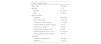
We analysed a total of 102 recordings from 86 patients, accounting for 8.5% of all EEG studies performed by the clinical neurophysiology department in the year 2020. Fifty-six patients (65.1%) were men and 30 (34.9%) were women. Mean age was 53 years (median, 60; range, 0-90). A total of 14 patients (17.4%) were aged under 14 years. Ninety-three percent of patients (n = 80) had history of one or more neurological diseases (Table 1).
Demographic characteristics, medical history, and medications used in the study population.
| Patient characteristics | |
|---|---|
| Mean age | 53 years |
| Gender | n (%) |
| Women | 30 (34.9) |
| Men | 56 (65.1) |
| Medical history | |
| Epilepsy | 28 (32.6) |
| Ischaemic stroke | 10 (11.6) |
| Intracranial haemorrhage | 11 (12.8) |
| Central nervous system surgery | 7 (8.1) |
| Intracranial infection | 4 (4.7) |
| Other origin cerebral lesion | 3 (3.5) |
| Severe traumatic brain injury | 47 (54.6) |
| Active SARS-CoV-2 infection | 2 (2.3) |
| Treatment | |
| Antiseizure medication | 65 (75.6) |
| Sedatives | 14 (16.3) |
The departments requesting ocEEG were the ICU (41.1%; n = 42), neurology (37.3%; n = 38), paediatrics (17.6%; n = 18), anaesthesiology (2%; n = 2), and neurosurgery (2%; n = 2).
EEG studies were performed in different areas of the hospital, depending on the indication and the patient’s clinical status. A total of 60 (58.8%) were performed in the ICU, including the paediatric (n = 12) and neonatal ICU (n = 4). The remaining studies were performed in the facilities of the clinical neurophysiology department; of these, 23.5% (n = 24) were from the emergency department and 17.6% (n = 18) were from conventional inpatient wards.
Mean study duration was 35.6 minutes (range, 20-123).
The main reason for ocEEG requests was suspected NCSE (56.9% of studies; n = 58), followed by monitoring of ASM or sedative treatment in patients with SE (21.6%; n = 22). Less frequent causes were evaluation of CRA (9.8%; n = 10), suspected brain death (6.9%; n = 7); new-onset ES (3.9; n = 4), and differential diagnosis between NEPE and ES (2.9%; n = 3). Fig. 2 summarises the indications and outcomes of ocEEG.

Distribution of the 102 EEG studies performed during on-call hours in 86 patients, classified by reason for the study, and the outcomes after interpretation of the results. It should be noted that EEG for treatment monitoring was performed in patients with non-convulsive status epilepticus, convulsive status epilepticus under treatment, or cardiorespiratory arrest with non-malignant EEG patterns and treatment-susceptible epileptic activity.
EEG: electroencephalography; ES: epileptic seizures; NEPE: non-epileptic paroxysmal events; SE: status epilepticus.
Fourteen patients (16.3%) underwent 2 or more EEG studies. With one exception, all of these were part of a single care process. The remaining patient underwent 2 EEG studies separated by an interval of several months, with both studies motivated by the same suspicion and confirming the diagnosis of NCSE.
Of the 58 patients undergoing ocEEG due to suspected NCSE, diagnosis was confirmed according to the Salzburg criteria17 in 20.7% of cases (n = 12); in the remaining patients, NCSE was ruled out. Fifty-two of these patients (89%) presented history of epilepsy or potentially epileptogenic brain injury.
Among the 22 patients in whom EEG was requested to monitor treatment response in SE, 22.7% (n = 5) showed persistent epileptiform discharges that continued to meet diagnostic criteria for SE; this result is helpful for adjusting treatment until resolution of ictal activity.
Seven patients underwent ocEEG in the context of CPA, whether to obtain prognostic information (10 recordings) or to diagnose and control seizures associated with cerebral anoxia. Of the 10 studies performed for prognostic assessment, 6 revealed malignant patterns, defined as suppressed background (< 10 µV), suppressed background with continuous periodic discharges, or burst suppression background with suppression periods constituting > 50% of the recording.18 In these cases, 100% of patients (n = 4) died. All 3 patients without malignant EEG patterns presented some degree of improvement.
In studies requested to support diagnostic suspicion of brain death, this diagnosis was confirmed in all cases (n = 7).
In 4 patients, emergency EEG was performed in the first hours after a new-onset ES. Recordings showed epileptiform abnormalities in 3 cases (75%), enabling diagnosis of epilepsy and supporting prescription of ASM.
In 3 patients presenting abnormal movements, EEG was requested to establish a differential diagnosis between ES and NEPE. Epileptic aetiology was confirmed in one patient, who met criteria for SE, and ruled out in the other 2.
Analysis of the anomalies identified in the recordings found that only 6.9% of recordings were considered normal (n = 7). The remaining 93.1% revealed some kind of alteration. The most frequent abnormality was interictal epileptiform activity (31.4%; n = 30), followed by diffuse slowing (30.4%; n = 29). Eighteen recordings (17.6%) were compatible with SE according to the Salzburg criteria.
Regarding treatment, no ASM onset or treatment escalation took place after EEG in 63% of cases. The largest group of patients was those with suspected NCSE that was not confirmed after EEG (n = 46), followed by those in whom ocEEG ruled out epileptic aetiology of abnormal movements (n = 3). These 2 groups account for 47.9% of all studies, and 56.3% of studies performed due to a suspected diagnosis with potential therapeutic implications (NCSE, new-onset ES, and differential diagnosis of ES vs NEPE). EEG monitoring of patients treated for SE detected persistence of ictal epileptiform abnormalities in 37% of cases (n = 8), supporting increased ASM treatment. Furthermore, ocEEG enabled diagnosis of epilepsy in 3 patients and confirmed epileptic aetiology in a patient with initially suspected NEPE. These patients account for 27.6% of all cases in which ocEEG had potential therapeutic implications (Fig. 1).
In some cases, ocEEG led to escalation of healthcare level, enabling optimal management of the disease diagnosed. Forty-two studies (41.2%) were performed in patients admitted to the emergency department or conventional inpatient wards. EEG results led to escalation of healthcare level in 22.2% of these cases (n = 9). The group of patients eligible for escalation of healthcare level included patients with suspected NCSE (confirmed in 6 cases), new-onset ES, or differential diagnosis between ES and NEPE. No escalation of healthcare level was observed among the patients undergoing EEG to monitor treatment response in EE, despite persistence of ictal epileptiform abnormalities (n = 2).
Results of ocEEG studies were available a mean of 31.6 hours (range, 6-139) earlier than would have been the case for routine EEG in ideal conditions (results available at 08:00 the next working day).
DiscussionWe present a series of patients who underwent ocEEG at the clinical neurophysiology department due to a range of indications, including life-threatening emergencies, emergencies with potential long-term functional repercussions, and, finally, situations in which essential information would be lost if it was not recorded immediately at the precise time at which they occur; this may take place outside of ordinary working hours.
On-call studies account for 8.5% of EEGs performed by the Hospital Universitario de Burgos clinical neurophysiology department in 2020. This is very similar to the rates reported in previous studies.6,19 It is important to note that these cases were gathered during the year 2020, when there was a reduction in outpatient EEG appointments due to the COVID-19 pandemic, resulting in an increase in the relative weight of emergency studies. The department performed 62, 67, and 90 ocEEG studies in the years 2017, 2018, and 2019, respectively.
In our series, the main reason for ocEEG was suspected NCSE, which accounted for 56.7% of all requests. This is consistent with the results reported by other authors.3,5,7,8 NCSE is a medical emergency with a broad range of differential diagnoses.20 It is diagnosed in up to 8% of patients with coma admitted to ICU,21 and is the underlying aetiological cause in 37% of patients assessed due to altered mental state.22 Diagnosis can only be confirmed or ruled out by an EEG study, as clinical data are insufficient and no alternative test providing this information is available. Delay in the diagnosis and treatment of NCSE and its underlying cause may result in poorer outcomes. In our series, ocEEG confirmed diagnosis in 20.7% of cases, ruling it out in the remaining 79.3%. These results enabled onset of appropriate treatment in the first group, and avoided unnecessary exposure to drugs in the second. When EEG is unavailable, all cases of suspected NCSE are treated empirically with ASMs, with a complete protocol in normal circumstances23; in a high percentage of patients, this treatment would have been unnecessary. In our series, 89% of patients in whom NCSE was confirmed presented history of potentially epileptogenic disease or an existing diagnosis of epilepsy (Table 1). In a retrospective series, Khan et al.9 identified 3 risk factors: prior history of epilepsy, witnessed seizure around the time of SE onset, and motor activity compatible with ES; NCSE was not observed in any patient with simultaneous absence of all 3 factors. However, the use of these criteria cannot be a substitute for ocEEG examination.
Diagnosis of CSE is a neurological emergency that does not typically require emergency EEG, and should be treated immediately. Nonetheless, DeLorenzo et al.10 report that at least 48% of patients with CSE in their series continued presenting ictal activity on EEG after resolution of motor activity; thus, EEG would be justified. Some authors recommend continuous EEG monitoring in patients with persistent altered mental state24; however, as this resource is unavailable at the majority of hospitals, we consider it appropriate to perform EEG monitoring after treatment onset, as this provides valuable information for treatment adjustment. In our series, ocEEG monitoring was performed in 22 patients after onset of treatment for SE (ASM or sedation). Persistence of ictal discharges was confirmed in 22.7% of these (n = 5); furthermore, it was possible to continue monitoring treatment/sedation until this activity resolved. Although, a priori, EEG diagnosis of NCSE is clearly defined by the Salzburg criteria, application of these criteria may be a challenge in patients in a coma and especially after onset of ASM/sedative treatment, leading to high levels of uncertainty. Understanding of patterns known as the ictal-interictal continuum and their careful interpretation by expert staff in the clinical context are essential to increasing diagnostic certainty,25,26 especially in patients admitted to the ICU, in whom misleading artefacts are frequently observed.
EEG provides both diagnostic and prognostic information after CRA, helping to adjust therapeutic effort. Despite the limited number of ocEEG studies performed in this context (n = 10), the patients presenting malignant patterns died, whereas all those patients without such patterns presented some degree of improvement. According to Westhall et al.,27 presence of various subcategories of malignant EEG pattern is highly sensitive and specific for prognostication of fatal outcome. This may result in a reduction in the duration of ICU stay in selected cases; in exceptional circumstances of healthcare system overload such as those seen in recent years, this may contribute to optimising resource use.
EEG continues to be relevant for diagnostic protocols for brain death. In our 7 patients undergoing ocEEG due to suspected brain death, this diagnosis was confirmed in all cases, contributing to the organ donation process. However, these patients only amounted to 6.9% of the total number of ocEEG studies performed. Had this been the only indication established for ocEEG, 93.1% of patients would have been excluded from the benefits the study provides for other indications.
Given the current definition of epilepsy, in which detection of epileptic anomalies on EEG may help to establish diagnosis13 and treatment onset, and the drop in the technique’s diagnostic yield within hours after the seizure,14,15 availability of emergency EEG is useful for both the diagnosis and the long-term management of epilepsy, helping to prevent seizure recurrence. The diagnostic yield of outpatient EEG studies is clearly inferior. In our series, only 4 patients underwent ocEEG for new-onset seizures; however, the results enabled a diagnosis of epilepsy to be established in 3 of these cases (75%).
Only 3 ocEEG studies were performed for differential diagnosis between NEPE and ES, with the study confirming SE in one of these and suggesting NEPE in the other 2; this contributes to the early onset of treatment after diagnosis, a fundamental aspect in the management of NEPE.28
The therapeutic implications of ocEEG studies may be classified into 2 groups: therapeutic decision-making, and adaptation of the level of healthcare to offer patients the most appropriate care for their needs. Some authors consider emergency EEG to be useful for diagnosis but not for therapeutic decision-making.7 According to our data, we avoided unnecessary ASM treatment in 79.3% of patients with suspected NCSE, in whom ocEEG ruled out this diagnosis (Fig. 1), and in 3 patients in whom epileptic aetiology was suspected but uncertain given the clinical signs observed. These cases with potential for iatrogenesis account for 47.9% of all studies and 56.3% of studies whose indication had therapeutic implications. It is reasonable to consider that avoiding unnecessary treatment would decrease morbidity and mortality associated with iatrogenesis; however, in addition to avoiding iatrogenesis, it is also important to provide appropriate treatment. In this regard, 37% of ocEEG studies performed to monitor treatment response in SE led to escalation of ASM treatment. Similarly, EEG enabled onset of treatment in 3 patients studied due to new-onset ES. Had ocEEG not been available, appropriate treatment would not have been provided in 11.7% of the total sample and 27.6% of studies conducted for indications with therapeutic implications. Furthermore, among patients admitted to the ICU, ocEEG performed to adjust treatment enables faster progression of patients, potentially reducing the mean duration of stays at a service characterised by highly specialised care.
With respect to decision-making regarding the appropriate healthcare level, ocEEG results supported escalation in 22.2% of patients who had not been admitted to the ICU. In the patients in whom this escalation did not take place, it is difficult to directly establish the usefulness of ocEEG, as these decisions are made on a multifactorial basis (ineligibility for ICU care due to the patient’s previous status, voluntary discharge, etc). However, with respect to the initial diagnostic suspicion, only 6 of the 29 cases of suspected NCSE were confirmed. Furthermore, among patients attended at the emergency department due to new-onset ES, diagnosis of epilepsy and onset of treatment enable studies to be performed on an outpatient basis; in patients undergoing EEG for differential diagnosis between ES and NEPE, only one case of SE was confirmed, with no epileptiform abnormalities observed in the rest of this group. In the absence of ocEEG, it is likely that all of these patients would have required hospital admission or escalation of healthcare level to manage the disease. It would be helpful for specific studies to assess the value of ocEEG in greater depth in this context.
In all the situations described above, our series suggests that availability of EEG outside of ordinary working hours enabled earlier diagnosis of the diseases for which the study was indicated, resulting in faster therapeutic decision-making. Therefore, diagnosis and treatment were established over a day earlier (mean, 31.6 hours) with respect to conventional EEG; this is fundamental in diseases with potentially severe outcomes, such as SE, but also prevents unnecessary exposure to the ASMs and sedatives used for the empirical treatment of such diseases as NCSE when these cannot be ruled out by ocEEG.
ConclusionsThe availability of EEG outside of ordinary working hours showed benefits in terms of diagnosis, treatment, and hospital management. It enables NCSE to be ruled out in a high percentage of suspected cases, preventing these patients’ exposure to a complete protocol of SE treatment, and justifies this treatment in those patients in whom diagnosis is confirmed. It helps adjust medication doses in SE under treatment, and enables treatment in patients who would not otherwise receive appropriate therapy. It enables diagnosis and therapeutic decision-making over 24 hours earlier than when EEG is performed exclusively during ordinary working hours. The results also inform decisions about escalation of healthcare level, optimising resource use, and may be highly valuable for the management of new-onset ES, differential diagnosis between ES and NEPE, and CRS occurring during on-call hours.
The efficiency of ocEEG in terms of diagnosis, treatment, and management justifies expanding timetables for its availability in hospitals.
LimitationsThis retrospective study has a small sample size, given the multiple, heterogeneous indications for ocEEG; this prevents us from drawing robust conclusions about its value for some of these diseases.
The authors have no conflicts of interest to declare.
