Buscar en
Medicina Clínica Práctica
Toda la web
Inicio Medicina Clínica Práctica Pneumatocele formation following COVID-19 pneumonia
Información de la revista
Vol. 6. Núm. 1.
(Enero - Marzo 2023)
Compartir
Compartir
Descargar PDF
Más opciones de artículo
Visitas
470
Vol. 6. Núm. 1.
(Enero - Marzo 2023)
Images in medicine
Open Access
Pneumatocele formation following COVID-19 pneumonia
FORMACIÓN DE NEUMATOCELE TRAS NEUMONÍA POR COVID-19
Visitas
470
Miguel Sogbea,b, I. Madeleine Di Friscoc, Ana Ezpondad, José Luis del Pozoa,e,f,
Autor para correspondencia
jdelpozo@unav.es

Corresponding author at: Infectious Diseases Division, Clínica Universidad de Navarra, Avda. Pio XII 36, 31008 Pamplona, Spain.
a Infectious Diseases Division, Clínica Universidad de Navarra, Pamplona, Spain
b Internal Medicine Department, Clínica Universidad de Navarra, Pamplona, Spain
c Pulmonary Medicine Department, Clínica Universidad de Navarra, Pamplona, Spain
d Radiology Department, Clínica Universidad de Navarra, Pamplona, Spain
e IdiSNA, Navarra Institute for Health Research, Pamplona, Spain
f Microbiology Department, Clínica Universidad de Navarra, Pamplona, Spain
Este artículo ha recibido

Under a Creative Commons license
Información del artículo
Texto completo
Descargar PDF
Estadísticas
Figuras (1)
Texto completo

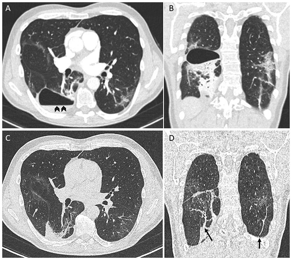
A 61-year-old man with no significant medical history presented to the emergency department with worsening dyspnea a week after close contact with someone who had COVID-19. He was unvaccinated. He was hypoxemic, and the chest radiograph showed bilateral opacities consistent with COVID-19 pneumonia and tested positive for RNA from SARS-CoV-2. Blood tests showed raised inflammatory markers. Computed tomography (CT) of the chest demonstrated bilateral ground-glass opacities. The patient was hospitalized and treated with high-flow nasal oxygen therapy, dexamethasone, and sarilumab. His clinical status improved, and he was discharged home after 1 week of hospitalization. Three weeks later, he presented again with worsening dyspnea, fever, and pleuritic chest pain. A CT pulmonary angiography ruled out pulmonary embolism (Fig. 1A, B) but demonstrated a thin-walled cystic lesion with an air–fluid level (Fig. 1A, arrowheads) that suggested an infected pneumatocele. The patient was managed conservatively with amoxicillin/clavulanic acid for 3 weeks. During the follow-up, the patient reported the disappearance of symptomatology. A subsequent CT performed 8 weeks after the diagnosis showed complete resolution of the pneumatocele (Fig. 1C, D) with persisting subpleural and parenchymal bands (Fig. 1D, arrows).

Fig. 1.

Chest Angio-CT. Thin-walled cystic lesion with an air-fluid level (arrowheads) (A) and consolidations in lower lobes (B). Chest Angio-CT performed eight weeks later showed complete resolution of the pneumatocele (C) with persisting subpleural and parenchymal bands (arrows) (D).

(0,18MB).
Funding

None.

Potential conflicts of interest

J.P. has been a consultant for Pfizer, MSD, Gilead, and Novartis. Other authors: No reported conflicts.

Acknowledgments

None

Copyright © 2022. The Authors
Opciones de artículo
Herramientas
es en pt

¿Es usted profesional sanitario apto para prescribir o dispensar medicamentos?

Are you a health professional able to prescribe or dispense drugs?

Você é um profissional de saúde habilitado a prescrever ou dispensar medicamentos