


Editado por:
José Antonio Sainz Bueno
Professor of Obstetrics and Gynecology, Faculty of Medicine, University of Seville, Seville, Spain Professor of Obstetrics and Gynecology, Faculty of Medicine, University of Seville, Seville, Spain Professor of Obstetrics and Gynecology, Faculty of Medicine, University of Seville, Seville, Spain Professor of Obstetrics and Gynecology, Faculty of Medicine, University of Seville, Seville, Spain Professor of Obstetrics and Gynecology, Faculty of Medicine, University of Seville, Seville, Spain
Eugenia Antolín Alvarado
Senior Consultant in Fetal Medicine, Department of Obstetrics and Ginecology, University La Paz Hospital; Member of the Obstetric Group in IdiPAZ- Biomedical Research Institute; Member of the SAMID network, Associate professor UAM University, Madrid, Spain
Última actualización: Julio 2025
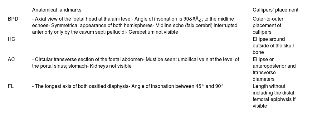
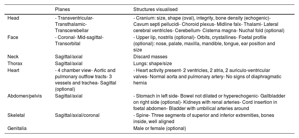
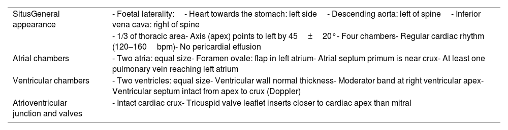
Más datosSecond trimester ultrasound is a standardised examination in pregnancy that should be routinely offered to all pregnant women, both for monitoring foetal growth and for screening for malformations. It should be performed between 18 and 24 weeks (in Spain from 18+0 to 22+0 weeks of gestation), by trained personnel and with appropriate equipment. The report should reflect foetal position and movements, biometry, amount of amniotic fluid, placental location and appearance, and foetal morphology. The foetal anatomy should include the study of the head (ossification and neurosonography), neck (discarding masses), thoracic cavity and its contents, abdomen and pelvis (studying stomach, umbilical vein, entrance of umbilical cord, kidneys, bladder), spine (in sagittal, coronal and axial planes), extremities (three segments, movement), and genitalia. Special attention should be paid to foetal heart examination (situs, four chamber view, left ventricular and right ventricular outflow-tracts, three-vessel and three-vessel-and trachea views). Neurosonography is also important with the transventricular, transcerebellar and transthalamic plane.
La ecografía del segundo trimestre de la gestación es una exploración estandarizada en el embarazo que debería ofrecerse de forma rutinaria a todas las gestantes, tanto para el seguimiento del crecimiento fetal como para el cribado de malformaciones. Debe realizarse entre las semanas 18-24 (en España de 18+0 a 22+0 semanas de gestación), por personal entrenado y con el equipamiento adecuado. El informe debe reflejar la posición y los movimientos fetales, la biometría, la cantidad de líquido amniótico, la localización y el aspecto de la placenta y la morfología fetal. La anatomía fetal debe incluir el estudio de la cabeza (osificación y neurosonografía), cuello (descartando masas), cavidad torácica y su contenido, abdomen y pelvis (estudiando estómago, vena umbilical, entrada del cordón umbilical, riñones, vejiga), columna vertebral (en planos sagital, coronal y axial), extremidades (3 segmentos, movimiento) y genitales. Debe prestarse especial atención al examen del corazón fetal (situs, vista de 4 cámaras, tractos de salida del ventrículo izquierdo y del ventrículo derecho, vistas de 3 vasos y de 3 vasos y tráquea). También es importante la neurosonografía con el plano transventricular, transcerebeloso y transtalámico.
