


Editado por:
José Antonio Sainz Bueno
Professor of Obstetrics and Gynecology, Faculty of Medicine, University of Seville, Seville, Spain Professor of Obstetrics and Gynecology, Faculty of Medicine, University of Seville, Seville, Spain Professor of Obstetrics and Gynecology, Faculty of Medicine, University of Seville, Seville, Spain Professor of Obstetrics and Gynecology, Faculty of Medicine, University of Seville, Seville, Spain Professor of Obstetrics and Gynecology, Faculty of Medicine, University of Seville, Seville, Spain
Eugenia Antolín Alvarado
Senior Consultant in Fetal Medicine, Department of Obstetrics and Ginecology, University La Paz Hospital; Member of the Obstetric Group in IdiPAZ- Biomedical Research Institute; Member of the SAMID network, Associate professor UAM University, Madrid, Spain
Última actualización: Julio 2025
Más datosEvaluation of the fetal heart is an integral part of prenatal care. Congenital heart defects (CHDs) are the most common major congenital anomalies, affecting approximately 0.8–1% of live births. Screening for CHDs has shown to improve perinatal outcomes. The performance of screening programs lies, among others, in the systematization of the sonographic evaluation. This review aims to outline the technique for conducting a basic fetal cardiac examination as well as to report the main indications for referral for advanced echocardiography and succinctly describe the technique for advanced echocardiography and first-trimester evaluation of the fetal heart.
La evaluación del corazón fetal es una parte integral de la atención prenatal. Los defectos cardiacos congénitos (CC) son las anomalías congénitas importantes más comunes y afectan aproximadamente al 0,8-1% de los nacidos vivos. Se ha demostrado que la detección prenatal de las malformaciones cardiacas mejora los resultados perinatales. El rendimiento de los programas de cribado prenatal de las anomalías cardiacas depende, entre otras cosas, de la sistematización de la evaluación ecográfica. Esta revisión tiene como objetivo describir: 1) la técnica para realizar un examen cardiaco, 2) las principales indicaciones para indicar la realización de una exploración avanzada o ecocardiografía, 3) la técnica para realizar la ecocardiografía y 4) la evaluación del corazón fetal en el primer trimestre.
Fetal echocardiography is an essential tool in prenatal care that enables a comprehensive assessment of both cardiac anatomy and function. Congenital heart defects (CHD) occur in approximately 8 out of every 1000 live births, making them the most common of severe congenital malformations and the ones that contribute most to infant mortality rates.1,2 Prenatal diagnosis improves pregnancy management, allowing a tailored follow-up, even offering prenatal therapy in selected cases, planning delivery logistics, and maximizing options for the family.3–7 Prenatal detection rates vary widely, based on maternal factors such as obesity or abdominal scars, fetal ones such as gestational age at evaluation or position, or external ones such as physician's experience, available equipment, or setting. A recent systematic review showed that prenatal identification of CHD remains disappointingly low, with many major defects being detected in below 50% of cases.8 However, it has been shown that resorting to a systematic approach in cardiac examination may increase detection rates.9–13
Evaluation of the fetal heart can be considered at two levels: a “basic” one targeting low-risk pregnancies and an “advanced” one, performed for those at higher risk for CHD. It is noteworthy that the majority of CHD (90%) occur in low-risk pregnancies and screening programs have shown a significant reduction in undiagnosed CHD at birth.14–16 Therefore, conducting a systematic and thorough examination of the entire population is crucial. This is optimally performed between 18 and 22 weeks’ gestation, although some anomalies can be identified from the first trimester and some will develop later on in pregnancy. Thus, current recommendations advocate for a stepwise approach in which the fetal heart should be evaluated in all scans with a special focus in the mid-trimester.17
The present review aims to describe the optimal sonographic settings as well as views required for a screening evaluation of the fetal heart as well as briefly describe the main indications for an advanced echocardiographic evaluation, additional views of the fetal heart, and particularities of the first trimester evaluation.
Ultrasound settings and technical factors for cardiac examinationA prerequisite for achieving an accurate diagnosis is the optimization of image quality in all sonographic modalities (M-mode, B-mode, pulsed Doppler, color Doppler). This implies making a series of basic adjustments, some of which are already preset in the different programs that the machines offer, to explore both the heart and the entire fetus throughout gestation. Initial adjustments for cardiac examination should be made in B-mode or grayscale, as other modalities are applied based from this image.18
The examination should be conducted transabdominally, reserving the transvaginal route for early gestational ages (12–16 weeks’ gestation) in high-risk patients for fetal heart conditions or situations where difficulties for second-trimester examination are anticipated (obese patients, abdominal scars, multifetal pregnancies, etc.).
Ultrasound transducerHigh-frequency probes improve the likelihood of detecting subtle defects. On the other hand, they have a reduced acoustic penetration which should be put into balance.
B-modeOptimization of the image should include maximizing the frame rate (number of images per second) by
- -
Adjusting the beam angle to the heart size and reducing the depth as much as possible.
- -
Using a single acoustic focus, placed at the lowest point of the area of interest.
- -
Activating zoom without incorporating color.
- -
Ensuring that the heart occupies between 1/3 and 1/2 of the screen.
- -
Increasing edge definition (with gain adjustment, low dynamic range, and use of harmonics).
Adjust the following for better quality:
- -
Window: adjust to focus on the area of interest, avoiding wide windows that reduce the number of images per second and subsequently grayscale image quality.
- -
Gain: start with lower gain and increase progressively until obtaining optimal imaging.
- -
PRF (pulse repetition frequency): tailor to the evaluated structures (high for high-speed flow such as auriculoventricular or semilunar valves and low for other vessels such as pulmonary veins).
- -
Filter: usually high, except for the examination of vessels with lower caliber to reduce interference from surrounding structures.
Key parameters for pulsed Doppler include:
- -
Incidence angle: avoid angles near 90°, as they result in poorly defined spectra or mirror images. Closer to 0° provides a more reliable velocity estimate.
- -
Sample volume: adapt it to the vessel size.
- -
PRF: adjust to the vessel speed to prevent aliasing or frequency overflow.
- -
Sweep speed: Higher in echocardiography compared to the study of other fetal organs.
Reserved for advanced echocardiography. Derived from the conventional B-mode image, TM mode does not visualize cardiac structures. Instead, each point along the axial axis is represented as a line moving over time. A fixed point results in a straight line, while moving points (ventricular walls, valves) create oscillating waveforms or sinusoids.
Cardiac exploration: views and techniqueThe focus of the current review lies on the basic or screening technique for cardiac examination. It consists of five axial planes described by Yagel et al. starting from the upper abdomen until the fetal mediastinum.19–22 They can be obtained by tilting the transducer toward the fetal head and include upper abdomen (situs), the four-chamber view (4CV), left ventricular outflow tract (LVOT), right ventricular outflow tract (RVOT) and the three vessel (3VV) or the three-vessel and trachea view (3VT). The addition of the outflow tracts as well as the 3VT to the 4CV increases diagnostic rates from 40–57% to 80–90%.23
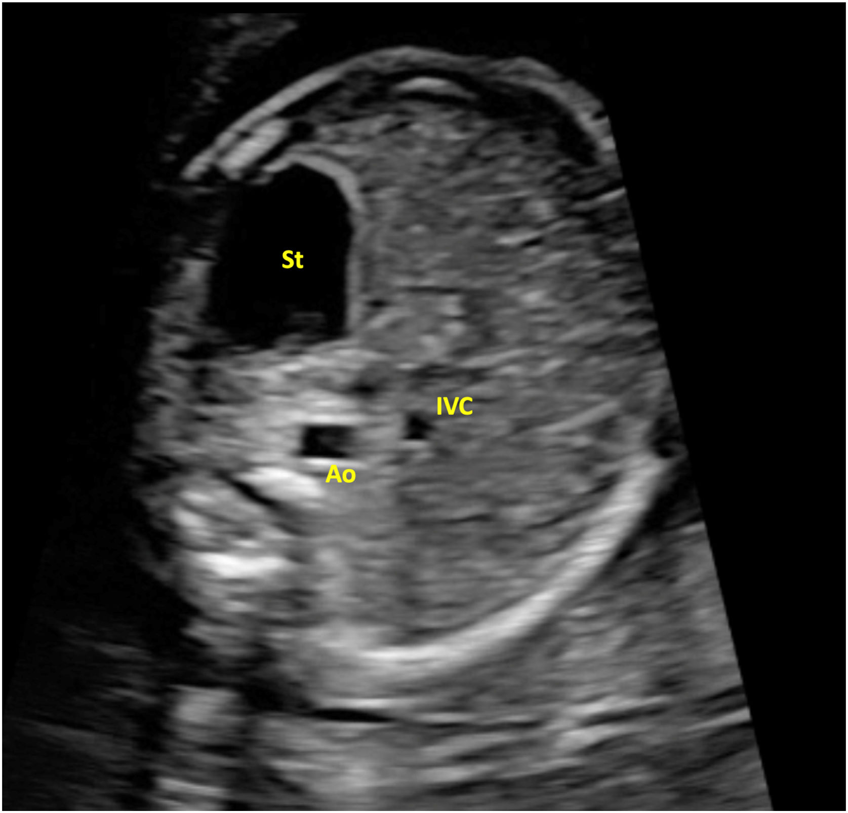
Upper abdomen viewAssessment of the cardiac and the visceral situs, which refers to the position of the heart/organs to the midline, requires first the identification of fetal laterality. The abdominal situs view is obtained at the level of the standard abdominal circumference measurement (below the fetal diaphragm) and a cephalad movement of the transducer to examine thoracic viscera and veno-atrial connections will allow evaluation of the cardiac situs.
The stomach should be located on the left side and cross-sectional views of the descending aorta and inferior vena cava should be identified on the left and right side of the spine, respectively (Fig. 1). Situs solitus is defined when these criteria are met, the vena cava drains into the right atrium and the heart is located on the left side of the thorax oriented to the left with an angle of 45±20° (levocardia, levoapex).
Four-chamber view
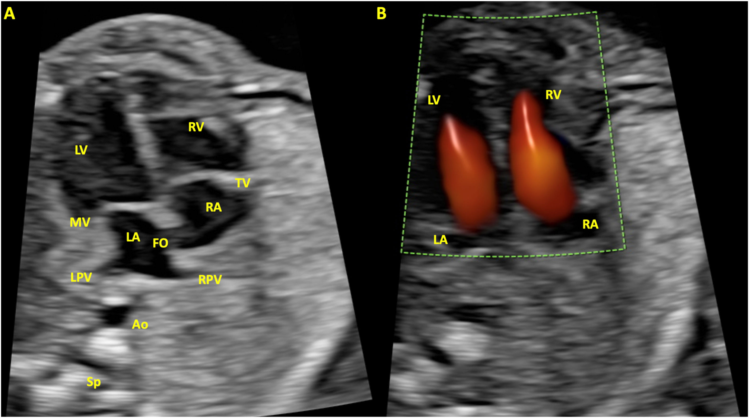
This is the most crucial and consistently used view in all cardiac examinations and can be achieved from two projections: apical or basal, where the ultrasound beam is parallel to the ventricular septum either entering from the apex (apical) or from the atria (basal), and subcostal or lateral where the beam is perpendicular to the ventricular septum. The most used view is the apical plane in which the following items must be evaluated (Fig. 2):
- -
Situs, cardiac position, and orientation: verify that the cardiac axis (the line extending the ventricular septum) forms an angle of 45±20° with a line crossing the fetal thorax from front to back. This results in approximately 2/3 of the heart on the left and 1/3 on the right.
- -
Cardiac size: considered normal if it occupies one-third of the total thoracic area, and the transverse diameter of the heart does not exceed 50% of the transverse diameter of the thorax.
- -
Presence of effusion: it is quite common to find a minimal amount of fluid in the pericardial sac, considered physiological if it is less than 2mm and does not exceed the level of the atrioventricular valves.
- -
Heart rate (normal between 120 and 160bpm) and subjective evaluation of cardiac contractility.
- -
Chamber symmetry: both atria and ventricles should be symmetrical. Although mild ventricular disproportion with right dominance can occur as a normal variant in the third trimester of pregnancy. However, marked right–left asymmetry should warrant further examination.
- -
Atria morphology: the left atrium is the most posterior structure, and at least two pulmonary veins should be visualized entering. The foramen ovale flaps within the left atrium. The right atrium has a more rounded morphology. The atrial septum primum is present (and is part of the crux cordis).
- -
Ventricular morphology: the left ventricle has smooth edges and only two papillary muscles, while the right one has three papillary muscles and a rougher appearance due to the moderator band, formed by the convergence of papillary muscles from the ventricular septum to the free wall at the most apical portion.
- -
Atrioventricular valves: insertion of both tricuspid and mitral annulus as well as their location should be identified. The septal leaflet of the tricuspid valve is located more apically and subsequently a portion of the ventricular septum will be shared between the left ventricle and the right atrium (so called atrioventricular septum).
- -
Ventricular septum: it can be divided into two portions, a larger muscular one representing two thirds of the septum and occupying the more apical part, and a membranous one occupying the proximal third. A sweep should be performed from the most posterior part of the septum toward the outflow tracts in a lateral view. Measurement of its thickness in this view allows assessment of normality and its deviations (hypertrophy).
- -
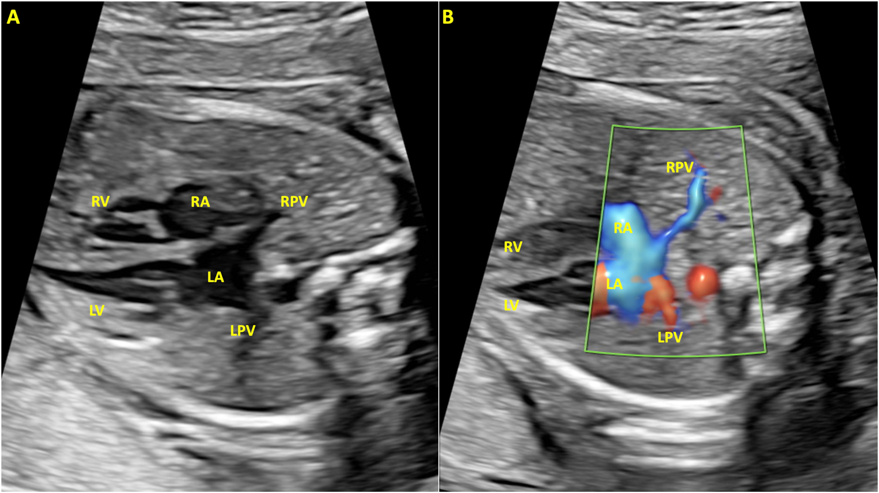
Doppler: routine color Doppler evaluation in the apical view is recommended to assess transvalvular flow. Pulsed Doppler evaluation of the atrioventricular valves should be included when suspecting anomalies with color Doppler to diagnose stenosis or insufficiency as well as disorders of cardiac rhythm. The caliper must be placed at the opening of the valves, in an apical projection, observing during diastole a biphasic wave with two cusps, called “e”, which corresponds to passive ventricular filling, and “a”, which corresponds to atrial contraction. Similarly, color Doppler in the subcostal view, lowering the PRF, enhance the examination of the pulmonary veins draining into the left atrium (Fig. 3).


Apical four chamber view. (A) B-mode, in systole, with both atrioventricular valves closed. (B) With color Doppler, in diastole, with the valves open. LV, left ventricle; RV, right ventricle; MV, mitral valve; TV, tricuspid valve; LA, left atrium; RA, right atrium; LPV, left pulmonary vein; RPV, right pulmonary vein; Ao, aorta; Sp, spine; FO, foramen ovale.
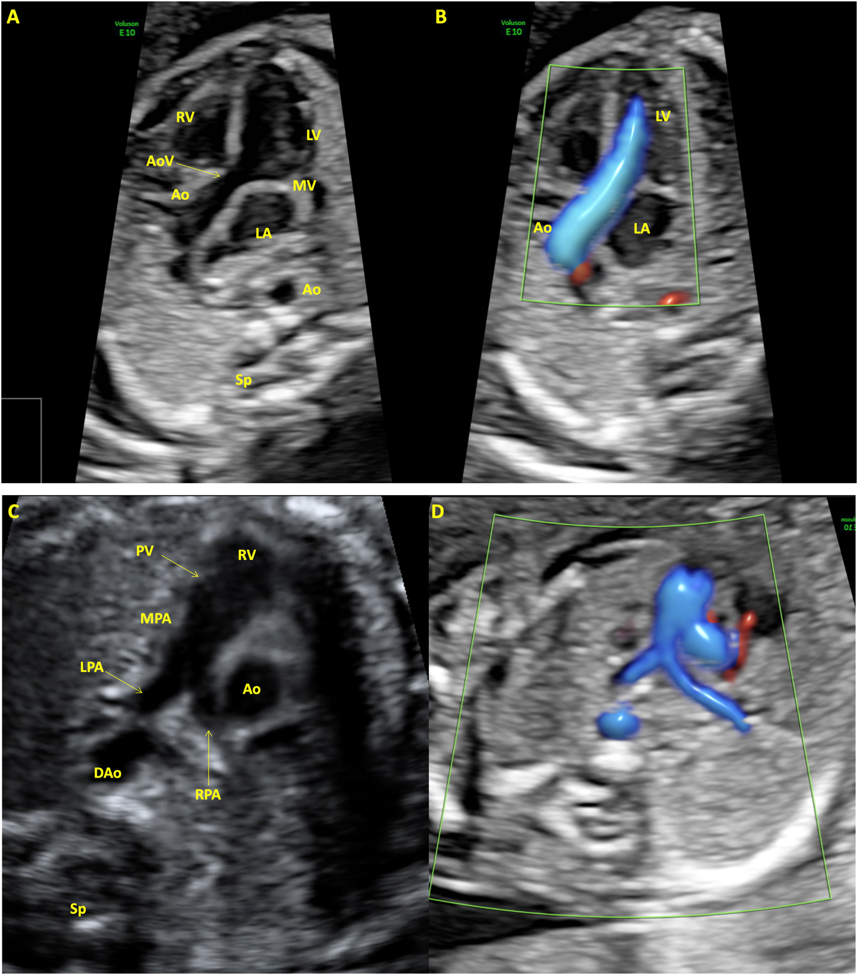
The aorta originates approximately in the center of the heart and shifts to the right. Therefore it is straightforward to generate a 5-chamber view of the LVOT from the previous 4CV (Fig. 4). A slight cephalad oscillation of the transducer will suffice. Alternatively, a rotational technique can be applied, especially from a lateral view, starting from the 4CV and rotating the transducer to the fetal right shoulder. It may require slightly more skill, but it optimizes the visualization of the LVOT, especially the outlet part of the septum that is in continuity with the anterior wall of the aorta. In either situation, it should be born in mind that it is only the presence of the supra-aortic vessels originating from it that confirms it as the aorta. The following items should be evaluated:
- -
Aortic outlet: as an extension of the ventricular septum. It allows evaluation of the integrity of the septum and excludes the existence of “outlet” septal defects.
- -
Mitro-aortic continuity: one side of the aortic valve appears as a continuation of the ventricular septum, while the other side appears next to the mitral valve annulus.
- -
Aortic valve: ensure that the diameter is appropriate, the valve opens completely in systole and closes normally in diastole, and transvalvular flow, examined with color Doppler, is laminar.

Left ventricular outflow tract view or five-chamber view in systole. (A) B-mode. (B) With color Doppler. The aortic valve is completely open and cannot be seen. Color Doppler demonstrates normal antegrade flow. Right ventricular outflow tract view in systole. (C) B-mode. (D) With color Doppler. The pulmonary valve is completely open and cannot be seen. LV, left ventricle; RV, right ventricle; MV, mitral valve; AoV, aortic valve; Ao, aorta; LA, left atrium; PV, pulmonary valve; MPA, main pulmonary artery; LPA, left pulmonary artery; RPA, right pulmonary artery; DAo, descending aorta; Sp, spine.
This view, obtained from the previous one by tilting the transducer toward the fetal head, enables visualization of the outlet of the pulmonary artery, originating from the central part of the right ventricle. It is slightly larger than the ascending aorta and crosses it anterior and cephalad to the LVOT almost at a right angle. The main pulmonary artery divides into two branches, right and left, and in the ductus arteriosus, which appears as the “extension” of the main vessel. Similarly to the LVOT, only the visualization of the two pulmonary branches identifies the vessel as the main pulmonary artery (Fig. 4). The following must be evaluated:
- -
Pulmonary artery outlet from the right ventricle.
- -
Crossing of the pulmonary artery over the aorta: the right outflow tract crosses over the left one in a perpendicular trajectory.
- -
Pulmonary valve: ensure that the diameter is appropriate, the valve opens in systole and closes normally in diastole and transvalvular flow, examined with color Doppler, is laminar.
This view is obtained at the level of the upper mediastinum, starting from the 4CV and moving the transducer upwards and slightly obliquely. Vessel number, size, alignment, and arrangement must be evaluated. From left to right, the pulmonary artery and ductus arteriosus, ascending aorta and aortic isthmus, and to the right of the aorta, the transverse section of the superior vena cava can be identified. Their relative diameters should decrease from left to right.
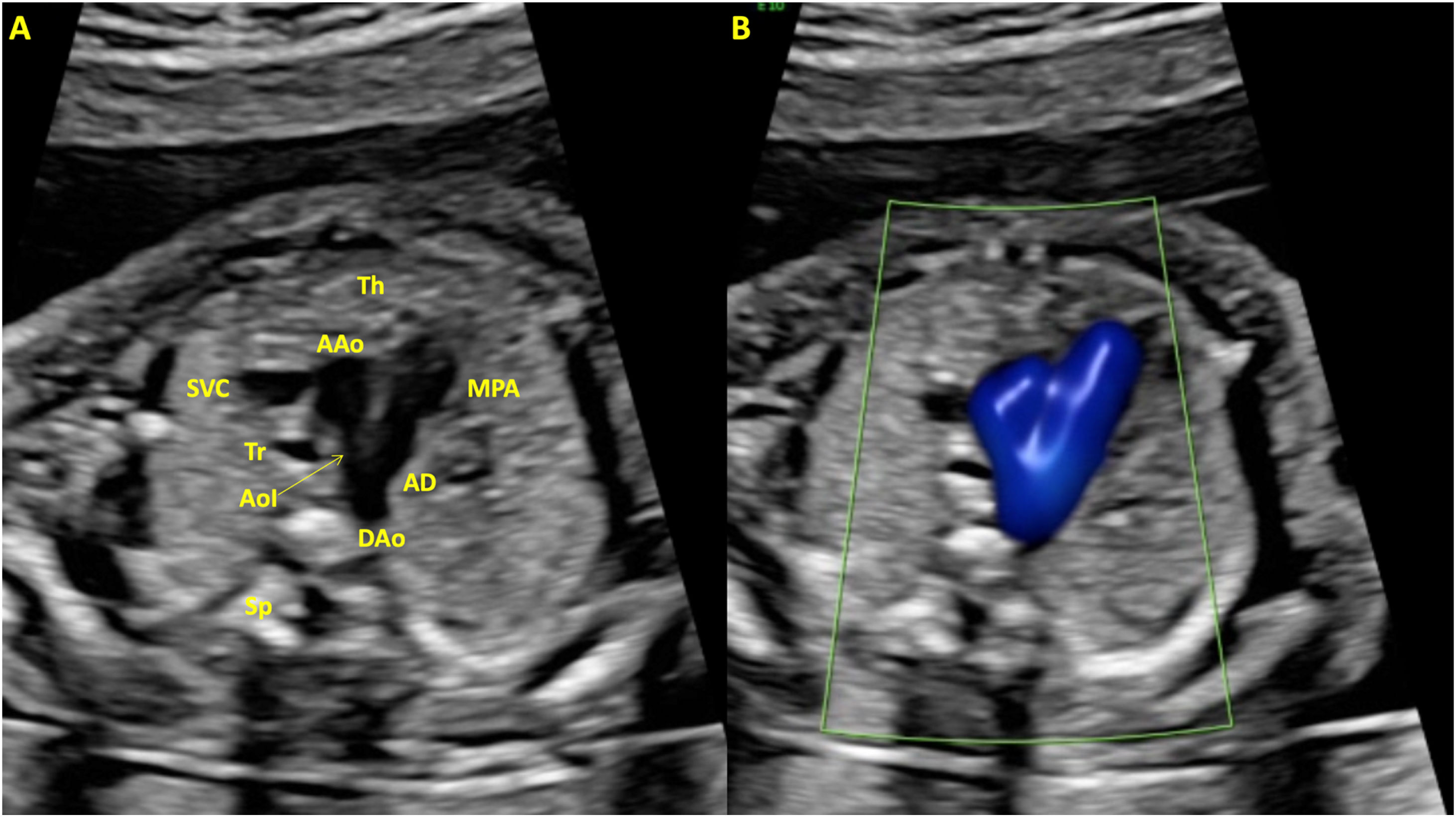
The 3VT view involves a more cephalad view from the prior one that includes the transverse aortic arch and the trachea, appearing in cross-section as a hyperechoic structure with fluid/anechoic content inside, located in front of the vertebral body and to the right of the aortic isthmus. The normal ductus arteriosus and left aortic arch course to the left of the trachea and converge forming an acute angle (‘V’-shape). The normal right superior vena cava and the normal thymus are also seen in this plane (Fig. 5).
Color Doppler
Recommendations about Doppler use during cardiac screening vary among guidelines being obligatory in some and highly recommended in others. Nevertheless, the ALARA use (as low as reasonably achievable) principle should be born in mind. Its use has proven useful for improving CHD detection rates especially in the setting of:
- -
Ventricular septal defects.
- -
Valvular abnormalities including stenosis, insufficiency, or atresia.
- -
Ductal-dependent CHD.
- 1)
The scan should start identifying fetal laterality, ideally locating presentation and the back.
- 2)
An axial plane of the upper abdomen (at the same location where the abdominal circumference is usually measured) should be obtained to start evaluating the visceral and cardiac situs.
- 3)
A cephalad sweep should be made until identifying the 4CV. Heart size and location should be assessed.
- 4)
At the 4CV both ventricles, atria and the cardiac apex can be visualized. The use of cine-loop is recommended. Assessment of the interventricular wall is preferably made from a lateral view.
- 5)
From the 4CV, perform a cranial sweep with little adjustment on the insonation angle to evaluate the crossover between the aorta and the main pulmonary artery in the LVOT. Alternatively, a rotational technique can be done twisting the probe toward the right fetal shoulder.
- 6)
Continue a cephalad movement to assess the RVOT.
- 7)
The 3VV and 3VT views are located cranial to the previous and discreetly oblique. Three vessels of decreasing diameter from left to right and the trachea should be visualized.
- 8)
Repeat the previous steps adding color Doppler and adjusting PRFs to the evaluated vessel.
Fetuses identified as having, or suspected of having, an abnormality on routine sonographic cardiac screening are candidates for advanced fetal echocardiography.24,25 The most frequent reasons for referral are listed below:
Fetal- -
High-risk aneuploidy screening without diagnostic test, suspicion of CHD during screening, or incomplete/suboptimal cardiac screening examination.
- -
Chromosomal defect.
- -
Nuchal translucency>99th centile (or ≥3.5mm) at 11–14 weeks’ gestation.
- -
Absent/reverse flow in ductus venosus between 11 and 14 weeks’ gestation.
- -
Suspected cardiac structural anomaly or arrhythmia (persistent tachycardia≥180bpm, bradycardia≤110bpm, or frequent irregular rhythm) on screening ultrasound.
- -
Extracardiac malformations associated with CHD.
- -
Non-immune fetal hydrops.
- -
Fetal growth restriction, especially when associated to other malformations.
- -
Fetal infection.
- -
Polyhydramnios, if there is an incomplete cardiac examination or if it is associated with other malformations.
- -
Monochorionic twin gestation.
- -
Anti-inflammatory treatment with non-steroidal anti-inflammatory drugs in the third trimester (high doses or continued).
- -
Pregestational diabetes.
- -
Anti-Ro/SSA antibodies.
- -
Phenylketonuria (with unknown periconceptional phenylalanine levels or >10mg/dL).
- -
Exposure to retinoids.
- -
First-degree relative with CHD (parents, siblings, half-siblings).
- -
First or second-degree relative with Mendelian inheritance disease with possible cardiac involvement.
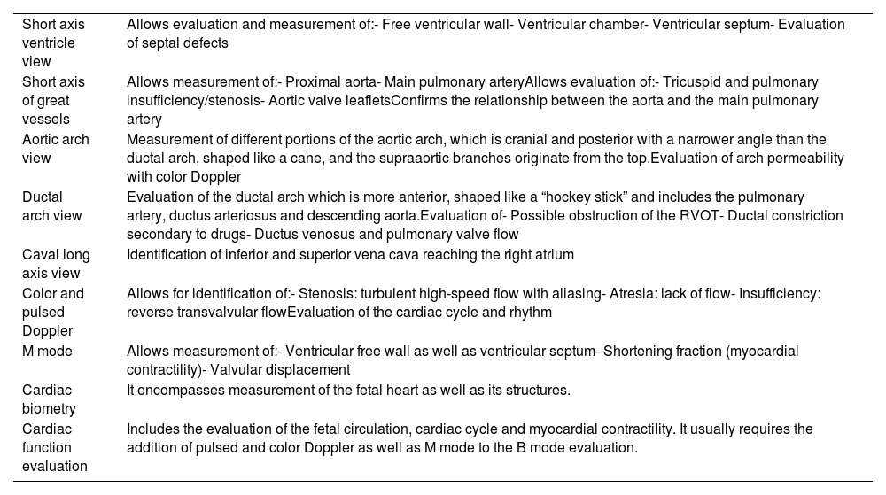
Advanced echocardiography includes the basic screening examination but can add other views to better assess cardiovascular structures (Fig. 5). Furthermore, other sonographic modalities that are not routinely employed can be of use. A brief summary of additional views and their use is presented in Table 1.
Accessory views and additional techniques included in advanced echocardiography.
| Short axis ventricle view | Allows evaluation and measurement of:- Free ventricular wall- Ventricular chamber- Ventricular septum- Evaluation of septal defects |
| Short axis of great vessels | Allows measurement of:- Proximal aorta- Main pulmonary arteryAllows evaluation of:- Tricuspid and pulmonary insufficiency/stenosis- Aortic valve leafletsConfirms the relationship between the aorta and the main pulmonary artery |
| Aortic arch view | Measurement of different portions of the aortic arch, which is cranial and posterior with a narrower angle than the ductal arch, shaped like a cane, and the supraaortic branches originate from the top.Evaluation of arch permeability with color Doppler |
| Ductal arch view | Evaluation of the ductal arch which is more anterior, shaped like a “hockey stick” and includes the pulmonary artery, ductus arteriosus and descending aorta.Evaluation of- Possible obstruction of the RVOT- Ductal constriction secondary to drugs- Ductus venosus and pulmonary valve flow |
| Caval long axis view | Identification of inferior and superior vena cava reaching the right atrium |
| Color and pulsed Doppler | Allows for identification of:- Stenosis: turbulent high-speed flow with aliasing- Atresia: lack of flow- Insufficiency: reverse transvalvular flowEvaluation of the cardiac cycle and rhythm |
| M mode | Allows measurement of:- Ventricular free wall as well as ventricular septum- Shortening fraction (myocardial contractility)- Valvular displacement |
| Cardiac biometry | It encompasses measurement of the fetal heart as well as its structures. |
| Cardiac function evaluation | Includes the evaluation of the fetal circulation, cardiac cycle and myocardial contractility. It usually requires the addition of pulsed and color Doppler as well as M mode to the B mode evaluation. |
There are additional techniques that can be applied to evaluate the fetal heart that are not as widely implemented as the prior ones. They are extrapolated from cardiac evaluation of the newborn and include, among others, speckle tracking analysis, 3D, 4D, and spatio temporal image correlation (4D STIC). They have proved to be of use in certain pathologies although their benefit over other more conventional approaches remains to be demonstrated.26,27
Cardiac biometryThe measurement of the structures of the fetal heart, especially the cardiac chambers, great vessels and valve annulus, is essential when facing a suspicion of a CHD. It allows to determine whether they are normal or not at a certain moment. Furthermore, they can be periodically reevaluated to obtain their growth curves and assess the progression of any cardiac defect.28 Two key issues must be accounted for in this process:
- -
The definition of normality throughout gestation.
- -
Systematize measurements appropriately to reduce intra- and inter-observer variability, ensuring that the results obtained are reproducible.
Regarding the first issue, normality can be defined either as a percentile or a z-score. There are many developed charts based on gestational age, femoral length or biparietal diameter. The chart used should always be referenced for better interpretability since the same measurement can be interpreted as normal or abnormal depending on the chosen reference standard.29–31
Structures should be measured at their maximum diameter. That is, global measurements of the heart, as well as those of the atrioventricular valves and cardiac chambers, should be performed in diastole whereas semilunar valves, great vessels, and their branches should be made in systole.
All measurements should be taken from inner edge to inner edge and the mean of three samples should be noted. Regarding ventricles, they should be measured in the 4CV, including the papillary muscles, tracing the endocardial contour parallel to the epicardium, and passing the lines at the level of the atrioventricular valves. For the ascending and descending aorta, as well as both cava veins, measurements are taken in a longitudinal plane. In the case of the pulmonary artery and its branches, an oblique section (short axis) or an oblique transverse plane of the fetal chest is used, showing the pulmonary artery in longitudinal view.32
Other relevant measurements include25:
- -
Cardiothoracic index (CTI): ratio of cardiac vs thoracic perimeter or area in a four-chamber section. Cardiomegaly is considered if the CTIp (perimeters) is greater than 0.55 or if the CTIa (areas) is greater than 0.35.
- -
Cardiac axis: The cardiac apex should be 45±20° to the left with respect to the anteroposterior axis of the thorax.
First-trimester examination of the fetal heart has substantially improved over the last two decades and screening at this gestational age has proved to be effective. Thus, most recent guidelines advocate for fetal cardiac screening in this period, although it is not mandatory, and its limitations must be acknowledged.33–36 Although visualization improves with advancing gestational age and the use of transvaginal ultrasound, it is not currently recommended to defer the scan, nor to routinely use the vaginal probe. Most guidelines agree that heart rate, situs, 4CV and 3VV/3VT should be evaluated, leaving the outflow tracts for an indirect evaluation either at the 3VV. Doppler should be used primarily for screening the 4CV and the 3VV. The following components are recommended as part of a detailed early cardiac screening examination:
- -
Situs: Identify the laterality of the stomach, heart, and cardiac axis.
- -
Four-chamber view: Use both B-mode and pulsed Doppler, taking advantage of the assessment of the tricuspid valve included in the screening for aneuploidies, ideally in an apical view.
- -
Three-vessel and trachea view: Use color Doppler to identify the laterality of the aortic and ductal arches.
- -
Due to the difficulty of visualizing the outflow tracts of the left and right ventricles, false positives and negatives are common. If CHD is suspected, the patient will be referred for an early advanced echocardiographic study (around 16 weeks’ gestation).
Fetal cardiology has evolved notably in the last three decades. However, there is still notorious dissociation between the high performance of fetal echocardiography, which is reserved for “high-risk” patients, and that of obstetric ultrasound with much poorer results. Although fetal echocardiography can diagnose prenatally most major CHD with a high degree of accuracy, enabling thereby appropriate parental counseling and perinatal management, every day clinical practice shows that many major CHD are still diagnosed postnatally in many centers despite of having been performed routine obstetric ultrasound scans. These poor results have been attributed classically to the difficulties in exploring the fetal heart. However, this is not absolutely true. Adherence to current recommendations on how to explore the fetal heart together with the proper skills of the examiner as well as the availability of a network of expert support to solve any suspicions, doubts or suboptimal scans are of paramount importance to increase the number of fetuses with major CHD who may benefit from prenatal diagnosis.
Ethical disclosuresProtection of human and animal subjectsThe authors declare that no experiments were performed on humans or animals for this research.
Confidentiality of dataThe authors declare that they have followed the protocols of their work center regarding the publication of patient data.
Right to privacy and informed consentThe authors declare that no patient data appear in this article.
FundingThis article is not funded.
Patient consentThe clinical management protocols of the H.U 12 de Octubre have been followed in the work and do not require patient consent for publication.
Conflict of interestThis article does not present any conflict of interest.



