


Editado por:
José Antonio Sainz Bueno
Professor of Obstetrics and Gynecology, Faculty of Medicine, University of Seville, Seville, Spain Professor of Obstetrics and Gynecology, Faculty of Medicine, University of Seville, Seville, Spain Professor of Obstetrics and Gynecology, Faculty of Medicine, University of Seville, Seville, Spain Professor of Obstetrics and Gynecology, Faculty of Medicine, University of Seville, Seville, Spain Professor of Obstetrics and Gynecology, Faculty of Medicine, University of Seville, Seville, Spain
Eugenia Antolín Alvarado
Senior Consultant in Fetal Medicine, Department of Obstetrics and Ginecology, University La Paz Hospital; Member of the Obstetric Group in IdiPAZ- Biomedical Research Institute; Member of the SAMID network, Associate professor UAM University, Madrid, Spain
Última actualización: Julio 2025
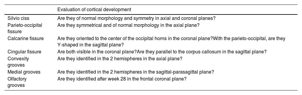
Más datosCongenital anomalies of the Central Nervous System are one of the most important and numerous groups of congenital malformations. They constitute the second cause of disability in childhood and more than 95% occur in a population without known risk. The most effective strategy for its detection is to differentiate between 2 levels of care. The first level, the BASIC ULTRASONOGRAPHY, is performed on all pregnant women, while the second level, the DETAILED FETAL NEUROSONOGRAPHY, is performed in cases selected due to the risk of anomaly based on a list of indications or because an abnormality has been detected or suspected. CNS abnormality on basic ultrasound. Its purpose is to perform a complete morphological and biometric multiplanar analysis of all accessible and recognizable brain structures from the axial, coronal and sagittal planes, ideally through transabdominal and transvaginal access. In the 3 plans, an attempt should be made to evaluate the same structures, assuming the limitations posed by the different perspective provided by each of them. When performing it, it is essential to take into account the indication and gestational age, know the morphological patterns, have reference tables of the normality of the different intracranial structures for GD and follow the systematics proposed by scientific societies. The objective of this article is to describe the different planes and provide readers with the key points for the ultrasound detection of the most important and frequent malformations of the CNS.
Las anomalías congénitas del Sistema Nervioso Central son uno de los grupos más importante y numeroso de las malformaciones congénitas. Constituyen la segunda causa de discapacidad en la infancia y más del 95% se presentan en población sin riesgo conocido. La estrategia más efectiva para su detección es diferenciar 2 niveles de atención. El primer nivel, la ECOGRAFÍA BÁSICA, se realiza a todas las gestantes, en tanto que el segundo nivel, la NEUROSONOGRAFÍA FETAL DETALLADA, se realiza en casos seleccionados por riesgo de anomalía a partir de un listado de indicaciones o por haber detectado o sospechado una anomalía del SNC en la ecografía básica. Su finalidad es realizar el análisis multiplanar morfológico y biométrico completo de todas las estructuras encefálicas accesibles y reconocibles a partir de los planos axiales, coronales y sagitales, idealmente por acceso transabdominal y transvaginal. En los 3 planos se debe tratar de evaluar las mismas estructuras, asumiendo las limitaciones que supone la diferente perspectiva facilitada por cada uno de ellos. Al realizarla es imprescindible tener en cuenta la indicación y la edad gestacional, conocer los patrones morfológicos, disponer de tablas de referencia de la normalidad de las diferentes estructuras intracraneales para la EG y seguir la sistemática propuesta por las sociedades científicas. El objetivo de este artículo es describir los diferentes planos y proporcionar a los lectores los puntos clave para la detección ecográfica de las malformaciones más importantes y frecuentes del SNC.
