


Analyze the etiology of infectious keratitis in our hospital.
Material and methodsRetrospective study in which the medical records of patients were reviewed in which a keratitis-producing microorganism was detected during the last 9 years (January 2014-December 2022). The sample was obtained by corneal scraping and seeded in non-selective media. Bacterial and fungal identification was carried out by mass spectrometry and viral identification by polymerase chain reaction (PCR). Sensitivity was obtained using disk-plate antibiograms, E-test or broth microdilution systems.
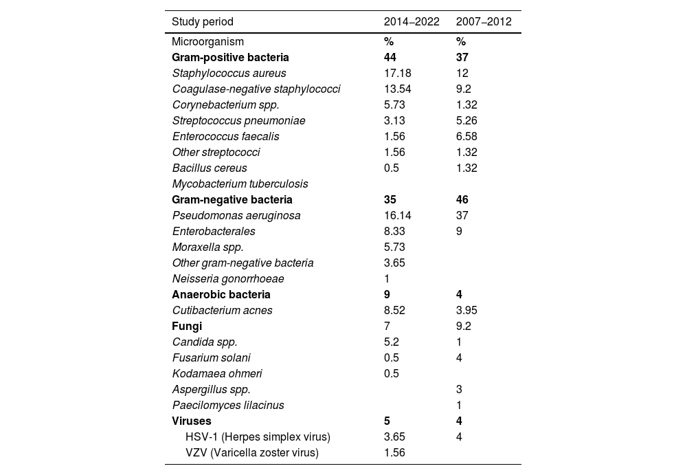
ResultsA total of 433 samples of corneal scrapings belonging to 416 patients were processed. Of the total samples, 196 were positive (44,3%). The average age was 55 years, with 51% being women. Regarding the etiology, we found the following isolates: Gram-positive bacteria (N=83) (44%), highlighting Staphylococcus aureus (N=33), coagulase-negative staphylococci (N=26), being Staphylococcus epidermidis the most frequent (N=19). Gram-negative bacteria (N=67) (35%), including: Pseudomonas aeruginosa (N=31), 42% associated with the use of contact lenses and Enterobacterales (N=16). Anaerobes (N=19), which 18 isolates were Cutibacterium acnes. Regarding viral etiology (N=10): herpes simple type 1 (N=7). Varicella-zoster virus (N=3). Finally, the fungal etiology (N=13), highlighting Candida spp. (N=10).
ConclusionsThe main agents of infectious keratitis are Staphylococcus aureus and Pseudomonas aeruginosa. The causative agent was detected in 44,3% of the samples, so microbiological analysis of these samples is highly advisable.
Analizar la etiología de la queratitis infecciosa en nuestro hospital.
Material y métodosEstudio retrospectivo en el que se revisaron las historias clínicas de pacientes en los que se detectó algún microorganismo productor de queratitis durante los últimos 9 años (Enero 2014- Diciembre 2022). La muestra se obtuvo mediante raspado corneal y se sembró en medios no selectivos. La identificación bacteriana y fúngica se realizó mediante espectometría de masas y la vírica por técnicas de reacción en cadena de la polimerasa (PCR). La sensibilidad se obtuvo mediante antibiogramas disco-placa, E-tests o sistemas de microdilución en caldo.
ResultadosSe procesaron un total de 433 muestras de raspados corneales pertenecientes a 416 pacientes. Del total de muestras, 196 fueron positivas (44,3%). La edad media fue 55 años, siendo el 51% mujeres. En cuanto a la etiología, encontramos los siguientes aislamientos: Bacterias grampositivas (N=83) (44%), destacando: Staphylococcus aureus (N=33), estafilococos coagulasa negativos (N=26), siendo Staphylococcus epidermidis (N=19) el más frecuente. Bacterias gramnegativas (N=67) (35%), entre ellas: Pseudomonas aeruginosa (N=31), un 42% asociados al uso de lentillas y Enterobacterales (N=16). Anaerobios (N=19), de los cuales 18 aislamientos fueron Cutibacterium acnes. En cuanto a la etiología vírica (N=10): herpes simple tipo 1 (N=7), virus varicela-zoster (N=3). Por último, la etiología fúngica (N=13), destacando Candida spp. (N=10).
ConclusionesLos principales agentes causales de la queratitis infecciosa son Staphylococcus aureus y Pseudomonas aeruginosa. En el 44,3% de las muestras estudiadas se detectó el agente causante por lo que el análisis microbiológico de estas muestras es muy aconsejable.
