Buscar en
Revista Española de Medicina Nuclear e Imagen Molecular (English Edition)
Toda la web
Inicio Revista Española de Medicina Nuclear e Imagen Molecular (English Edition) Visual and semi-quantitative analysis of 6-[18F]FDOPA PET/CT in patients with br...
Journal Information
Vol. 43. Issue 1.
Pages 6-13 (January - February 2024)
Share
Share
Download PDF
More article options
Visits
12
Vol. 43. Issue 1.
Pages 6-13 (January - February 2024)
Original Article
Visual and semi-quantitative analysis of 6-[18F]FDOPA PET/CT in patients with brain tumors and suspected tumor recurrence versus radionecrosis
Análisis visual y semicuantitativo del 6-[18F]FDOPA PET/TC en pacientes con tumores cerebrales ante la sospecha de recurrencia tumoral versus radionecrosis
Visits
12
M. Suárez-Piñeraa,b,
Corresponding author
marina.suarez.idi@gencat.cat

Corresponding author.
, L. Rodriguez-Bela, M. Alemanyb,c, A. Pons-Escodab,d, M. Pudisa, A. Coellob,e, G. Reynesf, N. Vidalb,g, M. Cortes-Romeraa, M. Maciab,h
a Unidad PET IDI, Servicio de Medicina Nuclear, Hospital Universitari de Bellvitge, L’Hospitalet de Llobregat, Barcelona, Spain
b Neuro-Oncology Functional Unit, Institut d'Investigació Biomèdica de Bellvitge-IDIBELL, L’Hospitalet de Llobregat, Barcelona, Spain
c Servicio de Neurología, Hospital Universitari de Bellvitge-ICO L'Hospitalet (IDIBELL), L’Hospitalet de Llobregat, Barcelona, Spain
d Servicio de Radiología, Sección de Neuroradiología, Hospital Universitari de Bellvitge, L’Hospitalet de Llobregat, Barcelona, Spain
e Servicio de Neurocirugía, Hospital Universitari de Bellvitge, L’Hospitalet de Llobregat, Barcelona, Spain
f Servicio de Física Médica, Hospital Universitari de Bellvitge-ICO L'Hospitalet (IDIBELL), L’Hospitalet de Llobregat, Barcelona, Spain
g Unidad de Neurooncología, Servicio de Anatomía Patológica, Hospital Universitari de Bellvitge, L’Hospitalet de Llobregat, Barcelona, Spain
h Servicio de Oncología Radioterápica, Institut Català d'Oncologia (ICO) L'Hospitalet (IDIBELL), L’Hospitalet de Llobregat, Barcelona, Spain
This item has received
Article information
Abstract
Full Text
Bibliography
Download PDF
Statistics
Figures (4)
Show moreShow less
Tables (5)
Table 1. Characteristics of the patients.
Table 2. Results obtained in a total of 70 lesions studied. There were 21 lesions in 20 patients, with primary brain tumors and 49 lesions in 42 patients with brain M1. Table 2 shows the results in the brain metastases.
Table 3. Shows the results in the primary brain tumors.
Table 4. Results of the diagnostic validity of joint visual and semi-quantitative analysis of PET/TC 6-[18F]FDOPA in patients with primary tumors and with brain metastasis and suspicion of tumor recurrence versus radionecrosis.
Table 5. Medians of the L/C and L/S ratios obtained in the semiquantitative analysis of PET/TC 6-[18F]FDOPA of tumor recurrence and radionecrosis in patients with primary brain tumors and brain metastasis.
Show moreShow less
Abstract
Introduction

Amino acid PET is a tool recommended by the main neuroimaging societies in the differential diagnosis between radionecrosis (RNC) and umour recurrence (TR) in brain tumours, but its use in our country is still limited. The aim of this work is to present our experience with 6-[18F]FDOPA PET/CT (FDOPA) in brain tumours (primary and M1), comparing these results with other published results.
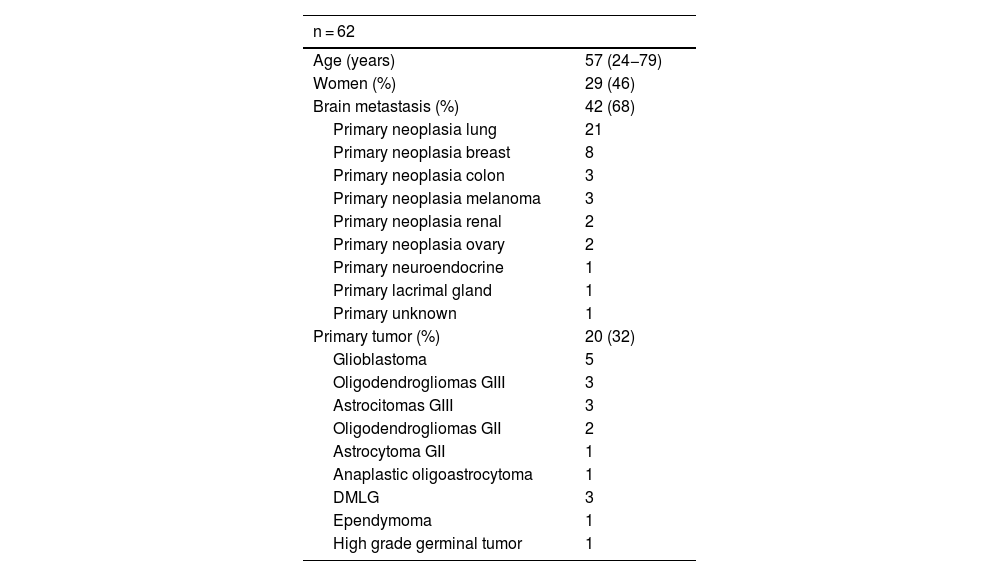
Material and methods

Retrospective study of 62 patients with suspected tumour recurrence (TR): 42 brain metastases (M1) and 20 primary, who underwent FDOPA. Images were analysed visually and semi-quantitatively, obtaining SUVmax and SUVmaxlesion/SUVmaxstriatum (L/S) and SUVmaxlesion/SUVmaxcortex (L/C) ratios. The diagnostic validity of PET was analysed and the best performing cut-off points were calculated. PET results were compared with clinical-radiological follow-up and/or histopathology.

Results

TR was identified in 49% of M1 and 76% of brain primaries. The best performing FDOPA interpretation was visual and semi-quantitative, with a sensitivity and specificity in primaries of 94% and 80% and in M1s of 96% and 72% respectively. The cut-off points with the best diagnostic performance were L/C1.44 in M1 and L/C1.55 in primaries. There are discrepant results with other published results.

Conclusion

FDOPA PET/CT is a useful tool in the differential diagnosis between recurrence and RNC in brain tumours. It is needed a standardization to contribute to homogenise FDOPA results a inter-centre level.

Keywords:
FDOPA
Radionecrosis
Tumores cerebrales
Estandarización
Resumen
Introducción

El PET con aminoácidos es una herramienta recomendada por las principales sociedades de neuroimagen, en el diagnóstico diferencial entre radionecrosis (RNC) y recurrencial tumoral (RT) en tumores cerebrales, sin embargo su uso en nuestro pais aún es limitado. El objetivo de este trabajo es presentar nuestra experiencia con 6-[18F]FDOPA PET/TC (FDOPA) en tumores cerebrales (primarios y M1), comparando estos resultados con otros publicados.

Material y métodos

Estudio retrospectivo de 62 pacientes con sospecha de recurrencia tumoral (RT): 42 metástasis cerebrales (M1) y 20 primarios, a los que se les realizó una FDOPA. Las imágenes fueron analizadas visual y semicuantitativamente, obteniendo el SUVmax y los ratios SUVmaxlesión/SUVmaxestriado (L/E) y SUVmaxlesión/SUVmaxcortex (L/C). Se analizó la validez diagnóstica del PET y se calcularon los puntos de corte con mayor rendimiento. Los resultados del PET se compararon con la evolución clínico-radiológica y/o con la histopatología.

Resultados

Se identificó RT en el 49% de las M1 y en el 76% de los primarios cerebrales. La interpretación de la FDOPA con mejores resultados fue la conjunta; visual y semicuantitativa, con una sensibilidad y especificidad en los primarios del 94 y 80% y en las M1 del 96 y 72% respectivamente. Los puntos de corte con mejor rendimiento diagnóstico fueron L/C1.44 en M1 y L/C1.55 en primarios. Existen resultados discrepantes con otros publicados.

Conclusión

La FDOPA PET/TC es una herramienta útil en el diagnóstico diferencial entre recurrencia y RNC en tumores cerebrales. Es necesario una estandarización que contribuya a homogeneizar los resultados de la FDOPA a nivel intercentro.

Palabras clave:
FDOPA
Radionecrosis
Brain tumour
Standardization

Article

These are the options to access the full texts of the publication Revista Española de Medicina Nuclear e Imagen Molecular (English Edition)
Subscriber
Subscriber

If you already have your login data, please click here .

If you have forgotten your password you can you can recover it by clicking here and selecting the option “I have forgotten my password”
Subscribe
Subscribe to

Revista Española de Medicina Nuclear e Imagen Molecular (English Edition)

Purchase
Purchase article

Purchasing article the PDF version will be downloaded

Price 19.34 €

Purchase now
Contact
Phone for subscriptions and reporting of errors
From Monday to Friday from 9 a.m. to 6 p.m. (GMT + 1) except for the months of July and August which will be from 9 a.m. to 3 p.m.
Calls from Spain
932 415 960
Calls from outside Spain
+34 932 415 960
E-mail
Article options
Tools
es en pt

¿Es usted profesional sanitario apto para prescribir o dispensar medicamentos?

Are you a health professional able to prescribe or dispense drugs?

Você é um profissional de saúde habilitado a prescrever ou dispensar medicamentos