


In lung cancer, axillary lymph node metastases (ALM) are rare, and according to the 8th grading system, it is classified as M1b disease. The aim of this study is to evaluate (i) the presence of ALM, and (ii) the effect of the primary tumors characteristics on the development of ALM.
MethodsWe performed a descriptive cross-sectional study, with retrospective revision, to identify ALM.
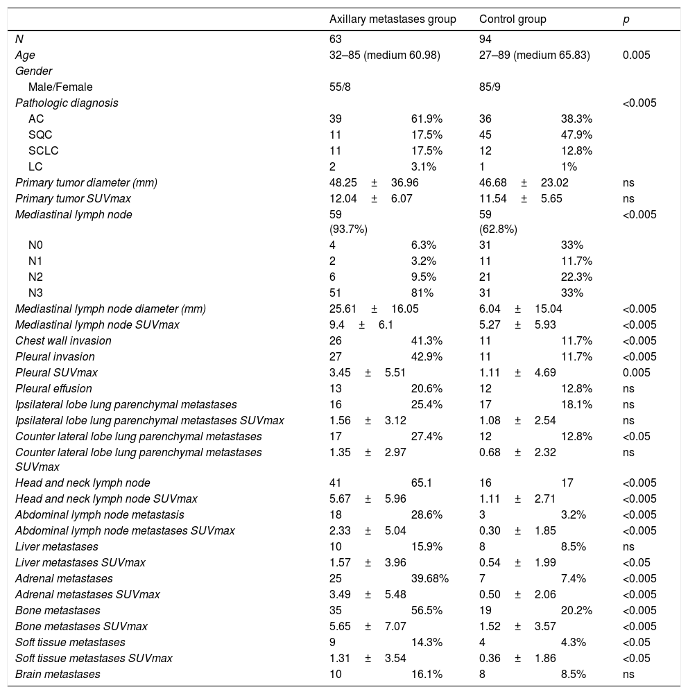
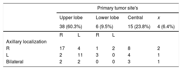
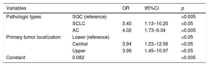
ResultsThere were 157 patients included in this analysis: ALM (63 patients) and control group (94 patients). The presence of extrathoracic lymph node, contralateral pulmonary parenchymal and distant metastasis and all SUVmax values were significantly higher in the study group versus the control group (p<0.05). The SUVmax value of the primary tumor was not a predictor of ALM. According to the primary histopathologic diagnosis, small cell lung cancer was found to cause ALM development 3.4 times as much as squamous cancer (SQC) (OR: 3.40 (95% CI 1.3–10.20), p=0.029) and adenocarcinoma group was found to cause ALM development 4 times as much as SQC (OR: 4.02 (95% CI 1.73–9.34), p=0.001). The likelihood of developing ALM was significantly higher in tumor located in the central and upper lobe versus the lower lobe.
ConclusionThe finding of ALM on PET/CT images, the necessity of histopathologic confirmation is determined according to the results of primary tumor localization, primary tumor histopathology, M stage on PET/CT imaging, localization of ALM according to primary tumor, and N stage on PET/CT imaging.
En el cáncer de pulmón, las metástasis en los ganglios axilares (ALM) son infrecuentes y, con arreglo a la 8ª edición sistema de estadificación, se clasifican como enfermedad metastásica M1b. El objetivo de este estudio es evaluar (i) la presencia de ALM, y (ii) el efecto de las características de los tumores primarios en el desarrollo de ALM.
MétodosRealizamos un estudio transversal descriptivo, con revisión retrospectiva, para identificar las ALM.
ResultadosIncluimos en este análisis a 157 pacientes: ALM (63 pacientes) y grupo control (94 pacientes). La presencia de ganglios extratorácicos, metástasis contralaterales pulmonares parenquimatosas y distantes, y todos los valores SUVmax fueron significativamente más elevados en el grupo de estudio, en comparación con el grupo control (p<0,05). El valor SUVmax del tumor primario no fue un factor predictivo de ALM. Con arreglo al diagnóstico histopatológico primario, detectamos que el cáncer de pulmón de células pequeñas causaba el desarrollo de ALM en una proporción 3,4 mayor que el cáncer de células escamosas (CCE) (OR: 3,4 (95% IC 1,3–10,2), p=0,029), y que el grupo de adenocarcinoma causaba el desarrollo de ALM en proporción 4 veces superior que el CCE (OR: 4,02 (95% IC 1,73–9,34), p=0,001). La probabilidad de desarrollar ALM fue considerablemente superior en los tumores localizados en el lóbulo superior que en el lóbulo inferior.
ConclusiónTras el hallazgo de ALM en las imágenes PET/TC, la necesidad de confirmación histopatológica viene determinada con arreglo a los resultados de la localización del tumor primario, la histopatología de éste, el estadio M en PET/TC, la localización de ALM con arreglo al tumor primario, y el estadio N en PET/TC.
Article
If you experience access problems, you can contact the SEMNIM Technical Secretariat by email at secretaria.tecnica@semnim.es or by phone at +34 619 594 780.

Revista Española de Medicina Nuclear e Imagen Molecular (English Edition)
