


To demonstrate the utility of 18F-FDG PET/CT in the differentiation of benign and malignant solitary fibrous tumors of the pleura (SFTP).
Materials and methodsA retrospective review was performed on the 18F-FDG PET/CT data from 17 patients with histopathologically diagnosed benign or malignant SFTP. The size, side of SFTP, presence of necrosis, calcification, pleural effusion, hilar lymphadenopathy (LAP), density on CT images (Hounsfield unit-HU), and 18F-FDG uptake (SUVmax) were recorded and compared in order to detect malignant SFTP. Statistical significance was set as p<0.05.
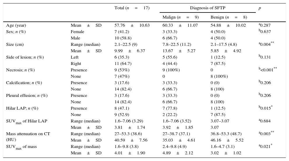
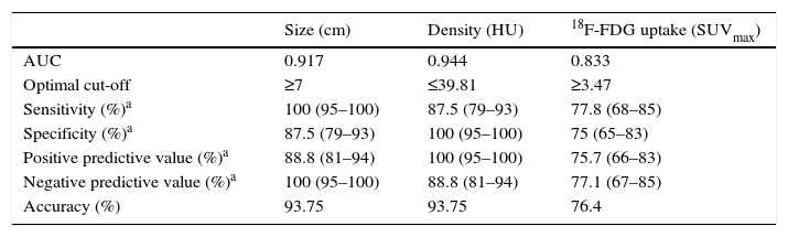
ResultsThe difference in size, presence of necrosis, and hilar LAP on CT images were statistically significant (p=0.004, p<0.001, p=0.015, respectively) in a comparison of benign and malignant SFTPs. The mean HU of benign SFTP was 46.16±5.52HU, and for malignant SFTP it was 35.03±4.61HU (p=0.003). The mean SUVmax was 3.02±1.02 for benign SFTP and 4.89±2.12 for malignant SFTP (p=0.021). A cut-off value of ≥7cm for size, ≤39.81HU for density, and ≥3.47 for SUVmax was obtained by ROC analysis for detecting malignant SFTP.
Conclusions18F-FDG PET/CT may have a limited role in diagnosing malignant SFTP in suspected patients.
Demostrar la utilidad de la 18F-FDG PET/TC en la diferenciación de benignidad o malignidad en tumores fibrosos solitarios de la pleura (TFSP).
Material y métodosSe revisaron retrospectivamente los datos de 18F-FDG PET/TC en 17 pacientes con diagnóstico histopatológico de TFSP benigno o maligno. Se valoraron el tamaño, la localización del TFSP, la presencia de necrosis, la calcificación, el derrame pleural, las adenopatías hiliares, la densidad de las imágenes de TC (unidades Hounsfield [HU]) y la captación de 18F-FDG (SUVmáx) para detectar TFSP maligno. La significación estadística se estableció como p<0,05.
ResultadosLa diferencia de tamaño, la presencia de necrosis y las adenopatías hiliares en imágenes de TC fueron estadísticamente significativas (p=0,004; p<0,001; p=0,015, respectivamente) comparando TFSP benignos y malignos. La media de HU en TFSP benigno fue 46,16±5,52 HU y en TFSP maligno fue 35,03±4,61 HU (p=0,003). El SUVmáx medio fue 3,02±1,02 para TFSP benigno y 4,89±2,12 para TFSP maligno (p=0,021). Un valor de corte ≥ 7cm de tamaño, ≤ 39,81 HU para la densidad y ≥ 3,47 para SUVmáx se obtuvo mediante análisis ROC para la detección de TFSP maligno.
ConclusionesLa 18F-FDG PET/TC tiene un papel limitado en el diagnóstico de pacientes con sospecha de SFTP maligno.
Article
If you experience access problems, you can contact the SEMNIM Technical Secretariat by email at secretaria.tecnica@semnim.es or by phone at +34 619 594 780.

Revista Española de Medicina Nuclear e Imagen Molecular (English Edition)
