


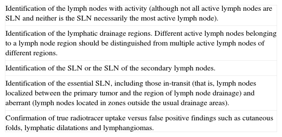
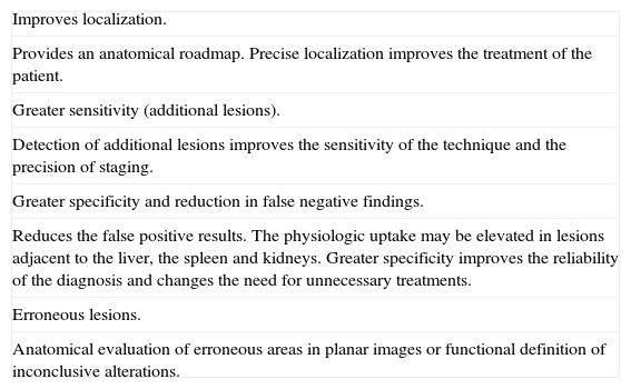
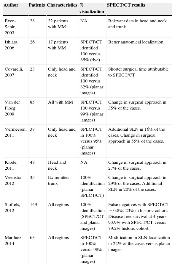
The main objectives of sentinel node (SN) biopsy are to avoid unnecessary lymphadenectomies and to identify the 20–25% of patients with occult regional metastatic involvement. This technique reduces the associated morbidity from lymphadenectomy and increases the occult lymphatic metastases identification rate by offering the pathologist the lymph nodes with the highest probability of containing metastatic cells. Pre-surgical lymphoscintigraphy is considered a “road map” to guide the surgeon toward the sentinel nodes and to localize unpredictable lymphatic drainage patterns. The SPECT/CT advantages include a better SN detection rate than planar images, the ability to detect SNs in difficult to interpret studies, better SN depiction, especially in sites closer to the injection site and better anatomic localization. These advantages may result in a change in the patient's clinical management both in melanoma and breast cancer. The correct SN evaluation by pathology implies a tumoral load stratification and further prognostic implication. The use of intraoperative imaging devices allows the surgeon a better surgical approach and precise SN localization. Several studies report the added value of such devices for more sentinel nodes excision and a complete monitoring of the whole procedure. New techniques, by using fluorescent or hybrid tracers, are currently being developed.
Los principales objetivos de la biopsia del ganglio centinela (GC) es evitar linfadenectomías innecesarias e identificar el 20–25% de pacientes que presentan enfermedad ganglionar regional clínicamente oculta. Esta técnica minimiza la morbilidad asociada a la linfadenectomía y aumenta también la tasa de identificación de metástasis linfáticas ocultas al ofrecer al patólogo aquel o aquellos ganglios con mayor probabilidad de contener células tumorales procedentes del tumor primario. La linfogammagrafía prequirúrgica se considera como un «mapa de carreteras» para guiar al cirujano hacia los GC y para la localización de patrones de drenaje impredecibles. Las ventajas del SPECT/TC incluyen un índice global de detección del GC superior a las imágenes planares, la capacidad para detectar ganglios centinelas en estudios convencionales difíciles de interpretar, mejor definición de los mismos en localizaciones cercanas a la inyección y una mejor localización anatómica. Estas ventajas pueden provocar un cambio en el manejo clínico-quirúrgico del paciente tanto en melanoma como en cáncer de mama. La correcta evaluación anatomopatológica del GC supone una estratificación de la carga tumoral y su posterior implicación pronóstica. La utilización de la imagen intraoperatoria permite al cirujano adaptar las marcas previamente hechas a la incisión quirúrgica planeada y confirmar la localización exacta del GC. Diversos estudios han demostrado el valor añadido de la utilización de estos dispositivos, al permitir la resección de GC adicionales y monitorizar el proceso quirúrgico. Nuevas técnicas, utilizando trazadores híbridos o fluorescentes, se están desarrollando en la actualidad.
Article
If you experience access problems, you can contact the SEMNIM Technical Secretariat by email at secretaria.tecnica@semnim.es or by phone at +34 619 594 780.

Revista Española de Medicina Nuclear e Imagen Molecular (English Edition)
