


Assess the role of SPECT-CT in sentinel lymph node (SLN) biopsy in the accurate anatomical location of the SNL in patients with cutaneous head and neck melanoma.
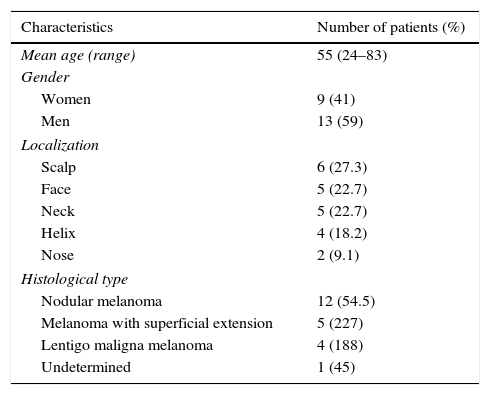
Material and methodsA retrospective study was conducted from February 2010 to June 2013 on 22 consecutive patients with a diagnosis of cutaneous head and neck melanoma (9 female, 13 male), with a mean age of 55 years old and who met the inclusion criteria for SLN biopsy. Patients underwent preoperative scanning after peri-scar injection of 99mTc-labeled-nanocolloid. Planar images of the injection-site, whole-body, and SPECT-CT scanning were acquired.
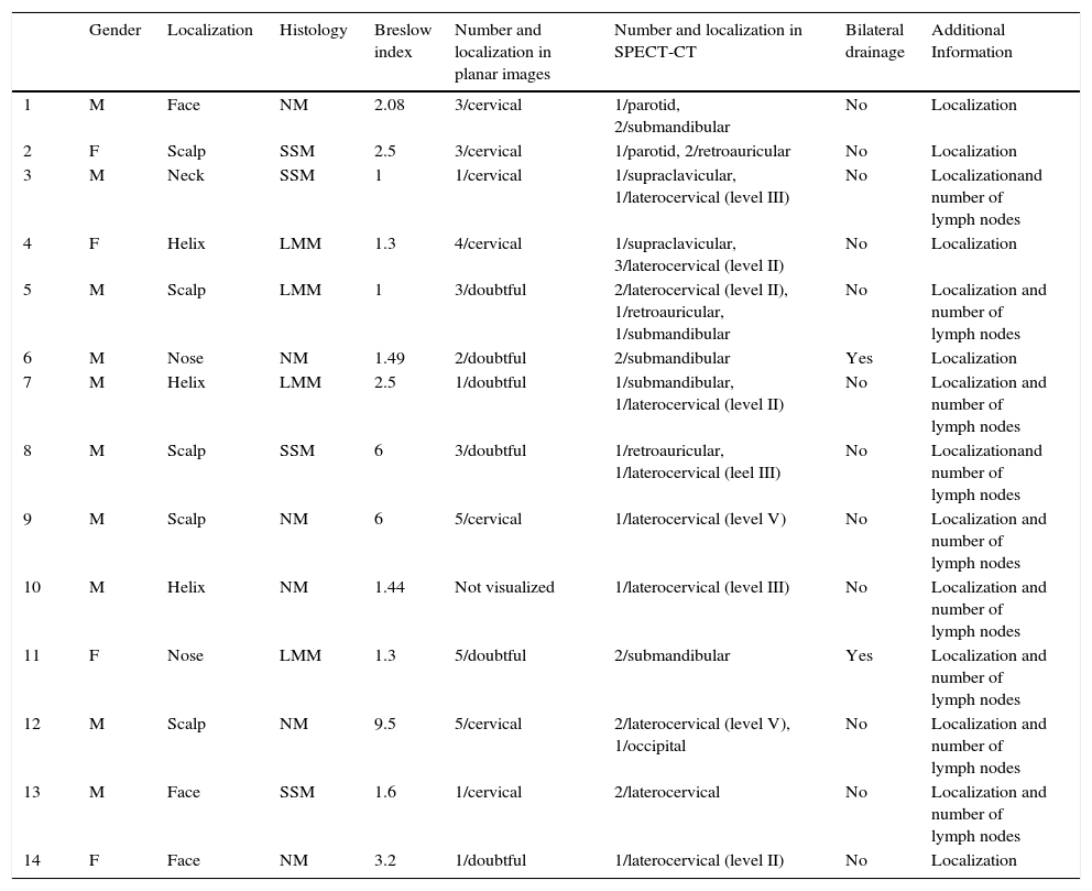
ResultsDetection rate of SLN reached up to 91% (20/22 patients) by planar lymphoscintigraphy and 95.4% (21/22 patients) by SPECT-CT. SPECT-CT provided an accurate location of SLN in 14/22 patients, enabling to improve the surgical approach (clinical impact: 63.6%). SLN was positive for metastatic cells in 9.1% patients.
ConclusionSPECT-CT provides detailed anatomical SLN location and allows detecting a higher number of SLN than planar lymphoscintigraphy. Routine use of SPECT-CT is recommended in order to optimize the SLN detection and location in patients with head and neck melanoma.
Valorar la aportación de la SPECT-TC a la biopsia selectiva del ganglio centinela (GC) en la correcta localización anatómica de este, en pacientes diagnosticados de melanoma cutáneo de cabeza y cuello.
Material y métodosEstudio retrospectivo entre febrero de 2010 y junio de 2013 que incluyó 22 pacientes consecutivos diagnosticados de melanoma cutáneo de cabeza y cuello (9 mujeres y 13 hombres), con una edad media de 55 años y criterios de inclusión de biopsia selectiva del GC. A todos ellos se les realizó una linfogammagrafía preoperatoria tras la inyección pericicatricial de nanocoloides de albúmina marcados con 99mTc, y posteriormente se obtuvieron imágenes planares sectoriales del lugar de inyección y de cuerpo completo, así como SPECT-TC.
ResultadosLa tasa de detección del GC fue del 91% (20 de 22 pacientes) para la linfogammagrafía planar y del 95,4% (21 de 22 pacientes) para SPECT-TC. En 14 de 22 pacientes la SPECT-TC mostró información relevante sobre la localización del GC modificando la vía de abordaje quirúrgica, siendo el impacto clínico de un 63,6%. En un 9,1% de los pacientes el GC fue positivo para metástasis de melanoma.
ConclusiónLa SPECT-TC proporciona información anatómica relevante sobre la localización del GC y detecta un mayor número de ganglios linfáticos que la linfogammagrafía. Se recomienda el uso rutinario de SPECT-TC en la linfogammagrafía del melanoma de cabeza y cuello para optimizar la localización y el número de GC en esta área.
Article
If you experience access problems, you can contact the SEMNIM Technical Secretariat by email at secretaria.tecnica@semnim.es or by phone at +34 619 594 780.

Revista Española de Medicina Nuclear e Imagen Molecular (English Edition)
