


The hypoxia-inducible factor 1 (HIF-1) has a critical role in oxygen homeostasis and it is a transcriptional activator of angiogenesis, erythropoiesis, iron and glucose metabolism. Glucose metabolism rate is increased in some tumours via HIF-1α. Our aim is to evaluate the relationship between hypoxia in colorectal cancer, PET parameters, necrotic tissue size and pathologic prognostic factors via using HIF-1α.
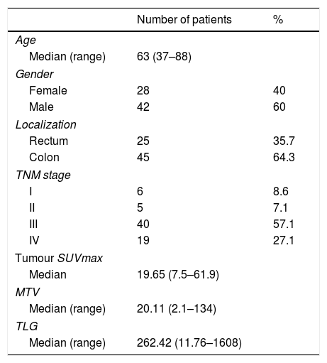
Materials/Methods70 patients (28female/42male; median age: 63 years) who were diagnosed with colorectal cancer via biopsy were staged with preoperative PET/CT and operated subsequently. Immunohistochemical evaluation scoring was done according to nuclear HIF-1α expression, staining density and intensity.
Metabolic tumour volume (MTV), total lesion glycolysis (TLG) and tumour volume (TV) were calculated by using volume of an ellipsoid formula via CT images, and percentage of tumour necrosis (%TmNcr) that was calculated by the difference between TV and recorded MTV.
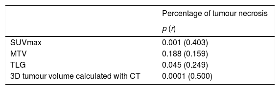
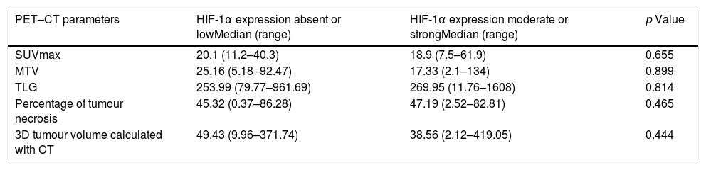
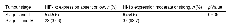
ResultsThere was a moderately meaningful positive correlation between tumour SUVmax and TV and %TmNcr (r=0.403, p=0.001 and r=0.500, p=0.0001, respectively). There were no statistically significant relationships between HIF-1α expression levels and tumour SUVmax, TLG, MTV, TV, %TmNcr, tumour stage, lymphovascular invasion, perineural invasion and extracapsular/capsular lymph node involvement. On the other hand, strong nuclear immunohistochemical staining was seen in tumour cells adjacent to invasive border, inflammatory cells. Although not statistically significant, moderate or strong nuclear staining were seen in 64.9% of metastatic patients.
ConclusionAlthough the presence of a positive correlation between tumour SUVmax and %TmNcr shows that there are hypoxic cells in cancer tissue with high FDG uptake, the relationship between the presence of HIF-1α and enhanced glucose metabolism and pathological prognostic factors of tumour was not shown. Strong nuclear immunohistochemical staining in tumour cells adjacent to invasive border and inflammatory cells leads us to believe that HIF-1α plays a role in the invasion area of tumour microenvironment.
El factor inducible para hipoxia (HIF-1) tiene un papel crítico en la homeostasis del oxígeno y es un activador transcripcional de angiogénesis, eritropoyesis, hierro y metabolismo de glucosa. La tasa de metabolismo de glucosa aumenta en algunos tumores a través de HIF-1α. Nuestro objetivo es evaluar la relación entre hipoxia en el cáncer colorrectal, los parámetros de PET, el tamaño del tejido necrótico y los factores pronósticos patológicos mediante el uso de HIF-1α.
Materiales/Métodos70 pacientes (28mujeres/42hombres; promedio de edad: 63 años) diagnosticados con cáncer co-lorrectal mediante biopsia, se estadificaron con PET/TC preoperatoria y se operaron posteriormente. La puntuación de evaluación inmunohistoquímica se realizó de acuerdo con la expresión de HIF-1α nuclear, la intensidad y la densidad de tinción. El volumen metabólico tumoral (MTV), la glucólisis de lesión total (TLG) y el volumen tumoral (TV) se calculó utilizando el volumen de una fórmula elipsoide mediante las imágenes de TC y el porcentaje de necrosis tumoral (%TmNcr) se calculó por diferencia entre TV y MTV.
ResultadosHubo una correlación positiva moderadamente significativa entre el SUVmax del tumor y TV y el %TmNcr (r=0,403,p=0,001 y r=0,5, p=0,0001,respectivamente). No hubo una relación estadísticamente significativa entre niveles de expresión de HIF-1α y SUVmax tumoral, TLG, MTV, TV,% Tm Ncr, estadio tumoral, invasión linfovascular, invasión perineural y afectación ganglionar extracapsular/capsular. Por otro lado, se observó una fuerte tinción inmunohistoquímica nuclear en las células tumorales adyacentes al borde invasivo, las células inflamatorias. Aunque no fue estadísticamente significativa, se observó una tinción nuclear moderada o fuerte en 64,9% de los pacientes metastásicos.
ConclusiónAunque la presencia de una correlación positiva entre SUVmax tumoral y el % de TmNcr muestra que hay células hipóxicas en tejido canceroso con una alta captación de FDG, no se demostró ninguna relación entre la presencia de HIF-1α y el incremento metabólico de glucosa y los factores patológicos del tumor. La fuerte tinción inmunohistoquímica nuclear en células tumorales adyacentes a las células inflamatorias y de borde invasivas nos hace pensar que HIF-1α desempeña un papel en el área de invasión del microambiente tumoral.
Article
If you experience access problems, you can contact the SEMNIM Technical Secretariat by email at secretaria.tecnica@semnim.es or by phone at +34 619 594 780.

Revista Española de Medicina Nuclear e Imagen Molecular (English Edition)
