


To evaluate the diagnostic yield of a selective brain 18F-FDG PET/CT in neurologically asymptomatic patients with small cell lung cancer.
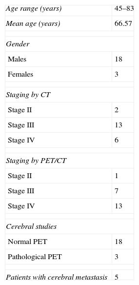
Material and methodsTwenty-one neurologically asymptomatic patients referred to our service between July 2008 and December 2009 for staging of small cell lung cancer were included in the study. All underwent a standard 18F-FDG PET/CT study followed by a selective brain PET/CT. The neurological findings were confirmed by CT scan with intravenous contrast, MRI or minimum clinical follow-up of 6 months.
The brain PET/CT was considered positive if any alteration was observed in the FDG distribution that was not related with previously known benign lesion in the CT image.
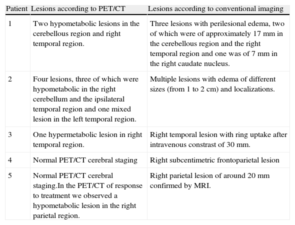
ResultsBrain metastases were detected in 5 of the 21 patients (23.8%), these being correctly classified in 3 of them by the selective brain PET/CT. The stage was upgraded in one of them with the selective brain study. Only one patient showed a hypermetabolic lesion in the PET images in relationship to the lesions observed in the CT images. Sensitivity, specificity, positive predictive value and negative predictive value were 60, 100, 100 and 88.89%, respectively.
ConclusionHypometabolic areas in the cerebral parenchyma are frequently associated to metastatic lesions in patients with small cell lung cancer. The selective brain PET/CT in these patients allows correct staging and early treatment of unsuspected metastasis.
Evaluar la rentabilidad diagnóstica del estudio selectivo cerebral con 18F-FDG-PET/TAC en pacientes con cáncer microcítico de pulmón asintomáticos neurológicamente.
Material y métodosSe incluyeron en el estudio 21 pacientes derivados a nuestro servicio entre julio de 2008 y diciembre de 2009, para estadificación, con histología de carcinoma microcítico de pulmón y asintomáticos neurológicamente. A todos ellos se les realizó un estudio 18F-FDG-PET/TAC estándar y a continuación un estudio selectivo cerebral, y se confirmaron los hallazgos neurológicos mediante TAC con contraste intravenoso, RM o seguimiento clínico mínimo de 6 meses. Un estudio cerebral PET fue considerado positivo si mostraba cualquier alteración en la distribución de la FDG no relacionada con lesiones benignas previas en la TAC cerebral.
ResultadosEn 5 de los 21 pacientes (23,8%) se detectaron metástasis cerebrales, siendo correctamente diagnosticados mediante 18F-FDG-PET/TAC 3 de ellos. En uno de ellos la realización del estudio cerebral incrementó el estadio. Sólo uno de los pacientes mostró hipermetabolismo en la imagen PET en relación con las lesiones cerebrales evidenciadas en la imagen TAC. Se obtuvieron valores de sensibilidad, especificidad, valores predictivos positivo y negativo del 60, 100, 100 y 88,89%, respectivamente.
ConclusiónLas áreas hipometabólicas en el parénquima cerebral con frecuencia se asocian a lesiones metastásicas en pacientes con cáncer microcítico de pulmón. La realización de un estudio selectivo cerebral PET/TAC en estos pacientes permite una correcta estadificación y el tratamiento precoz de las metástasis no sospechadas.
Article
If you experience access problems, you can contact the SEMNIM Technical Secretariat by email at secretaria.tecnica@semnim.es or by phone at +34 619 594 780.

Revista Española de Medicina Nuclear e Imagen Molecular (English Edition)
