


To analyze the potential improvement of 18F-fluorodeoxyglucose (FDG) PET/CT using additional delayed images of the liver in operated colorectal cancer.
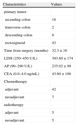
Material and methodsThe study prospectively included 71 patients (22 women, 49 men) with mean age of 65±11 years with clinical, biochemical or radiological suspicion of current disease. A whole body PET/CT scan was performed at 60min (standard images) and after 2h (delayed images) post-injection of 4.07MBq/kg of 18F-FDG. Visual and quantitative SUV analysis of PET/CT findings were done. All findings were confirmed by histopathology and/or at least 6 months follow-up.
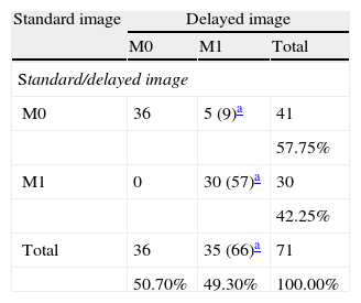
ResultsThirty-seven out of 71 patients were diagnosed of liver metastases (79 metastases). In 38/71 cases there was extra-hepatic disease in the form of local recurrence (10), abdominopelvic (3) or mediastinal (3) lymph nodes, bone (1) or lung metastases (16) and carcinomatosis (10). Sensitivity and specificity for the diagnosis of liver metastases in a patient-by-patient basis in standard (81% and 91%) and in delayed images (95% and 97%) were calculated. The number of lesions detected in delayed images was significantly higher (66/79) than in standard images (57/79). Sensitivity and specificity for PET/CT in the diagnosis of extra-hepatic disease were 84% and 70%, contributing to the detection of synchronous tumors in 5 patients.
ConclusionsPET/CT may be useful in the diagnosis of extra-hepatic disease in suspected recurrence of colorectal cancer. Delayed images on PET/CT may increase the sensitivity to identify liver metastases.
Analizar el potencial de la PET/TC usando imagen tardía del hígado en pacientes con sospecha de recidiva de cáncer colorrectal.
Material y métodosSe han incluido prospectivamente 71 pacientes (22 mujeres, 49 hombres) con edad de 65±11 años y sospecha clínica, analítica o radiológica de recurrencia. Se realizó PET/TC después de la inyección de 4,07MBq/kg de 18F-FDG con imagen de cuerpo entero a los 60min (imagen estándar) y hepática a las 2h (imagen tardía). Se efectuó análisis visual y cuantitativo mediante SUV de los hallazgos de la PET/TC. Se obtuvo confirmación de las lesiones por estudio histopatológico y/o seguimiento mínimo de 6 meses.
ResultadosSe diagnosticaron metástasis hepáticas en 37/71 pacientes (79 metástasis). Un total de 38/71 pacientes mostraban enfermedad extra-hepática en forma de recidiva local (10), adenopatías abdominopélvicas (3) o mediastínicas (3), metástasis óseas (1) o pulmonares (16) y carcinomatosis (10). Se calculó la sensibilidad y especificidad para el diagnóstico de metástasis hepáticas en base a cada paciente para la imagen estándar (81 y 91%) y la imagen tardía (95 y 97%). El número de metástasis hepáticas diagnosticadas fue mayor con la imagen tardía (66/79) que con la imagen estándar (57/79). La sensibilidad y especificidad de la PET/TC en lesiones extra-hepáticas fue de 84 y 70%, contribuyendo al diagnóstico no sospechado de 5 tumores sincrónicos.
ConclusionesLa PET/TC es recomendable para descartar enfermedad extra-hepática en sospecha de recidiva de cáncer colorrectal. La realización de imagen tardía mejora la sensibilidad de la PET/TC en el diagnóstico de metástasis hepáticas.
Article
If you experience access problems, you can contact the SEMNIM Technical Secretariat by email at secretaria.tecnica@semnim.es or by phone at +34 619 594 780.

Revista Española de Medicina Nuclear e Imagen Molecular (English Edition)
