


We aimed to evaluate the diagnostic impact of 18F-FDG PET/CT in staging apocrine breast carcinoma (ABC) and primary breast neuroendocrine carcinoma (PBNEC) and to demonstrate possible alterations of the 18F-FDG uptake in these histopathologic subtypes. In addition, we aimed to compare 18F-FDG PET/CT findings between ABC, PBNEC and invasive ductal carcinoma (IDC).
Material and methodsA total of 570 patients and 585 breast lesions were retrospectively included in this study. After patients were classified into molecular subtypes according to the histopathological analysis, 18F-FDG PET/CT imaging was performed. The SUVmax findings of primary tumors obtained from 18F-FDG PET/CT were compared between the groups.
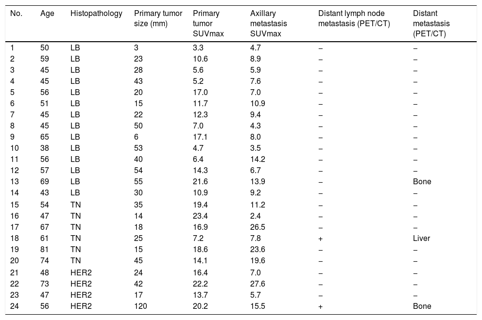
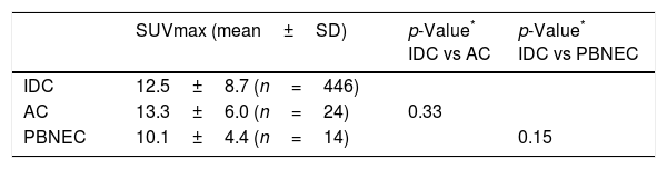
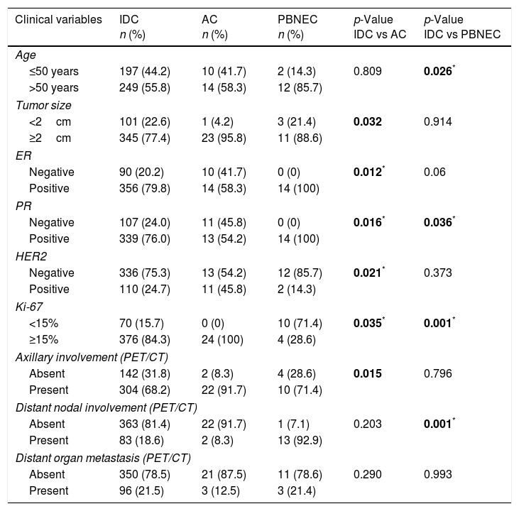
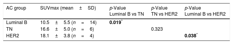
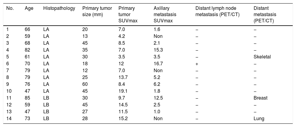
ResultsIDC was the most prevalent breast carcinoma (77.7%, n=446), with a low proportion of ABC (4.1%, n=24) and PBNEC (2.4%; n=14) diagnosed. The highest mean SUVmax was calculated in HER2 subtype of ABC and 18F-FDG uptake ratio in HER2 and TN subtypes were found statistically higher than Luminal B type of ABC (p=0.038 and p=0.019, respectively). Although 18F-FDG uptake in Luminal B subtype of PBNEC was higher than Luminal A subtype, difference was not statistically significant. Additionally, the axillary metastasis rate was significantly higher in the ABC group (p=0.015).
ConclusionsThe histopathological ABC subtype group showed different 18F-FDG uptake than the IDC group. Even if 18F-FDG uptake was lower in the PBNEC group than in the other groups, PET/CT showed and adequate performance in detecting primary tumors and metastases. The 18F-FDG PET/CT scan results may contribute to the initial staging and management of ABC and PBNEC patients.
El objetivo fue evaluar el impacto diagnóstico y la estadificación mediante PET/TC con 18F-FDG en carcinomas apocrinos (ABC) y carcinomas neuroendocrinos (PBNEC) y detectar posibles alteraciones en la captación de 18F-FDG según el subtipo histológico de estos tumores. Además, nuestro objetivo fue comparar los hallazgos de la PET/TC con 18F-FDG entre ABC, PBNEC y el carcinoma ductal invasivo (IDC) de mama.
MétodosSe incluyeron restrospectivamente un total de 570 pacientes y 585 lesiones mamarias. Después de clasificar a los pacientes en subtipos moleculares de acuerdo con el análisis histopatológico, se realizaron imágenes de PET/TC con 18F-FDG y se compararon los hallazgos del análisis visual y SUVmax de los tumores primarios entre los distintos grupos.
ResultadosEl IDC fue el carcinoma de mayor prevalencia, con una tasa de detección del 77.7% (n= 446) en nuestro estudio. Sin embargo, se diagnosticaron 4.1% (n= 24) ABC y 2.4% (n= 14) PBNEC. El SUVmáx medio más alto se registró en el subtipo HER2 del ABC y se encontró una diferencia estadísticamente significativa entre la ratio de captación de 18F-FDG de los subtipos HER2 y TN comparados con el tipo luminal B del ABC (p= 0.038, p= 0.019, respectivamente). Aunque la captación de 18F-FDG en el subtipo Luminal B de PBNEC fue mayor que el subtipo Luminal A, la diferencia no fue estadísticamente significativa.Además, la tasa de metástasis axilar fue significativamente mayor en el grupo ABC que en los otros grupos (p= 0.015).
ConclusionesLos subtipos histopatológicos del grupo ABC mostraron diferentes grados de captación de 18F-FDG con respecto a los subtipos del grupo IDC, a pesar de que en general la captación de 18F-FDG fue menor en el grupo de PBNEC. La PET/TC demostró ser adecuada en la detección de tumores primarios y metástasis en nuestro estudio. Los resultados de la exploración PET/TC con 18F-FDG pueden contribuir a la estadificación inicial y al manejo de pacientes con ABC y PBNEC.
Article
If you experience access problems, you can contact the SEMNIM Technical Secretariat by email at secretaria.tecnica@semnim.es or by phone at +34 619 594 780.

Revista Española de Medicina Nuclear e Imagen Molecular (English Edition)
