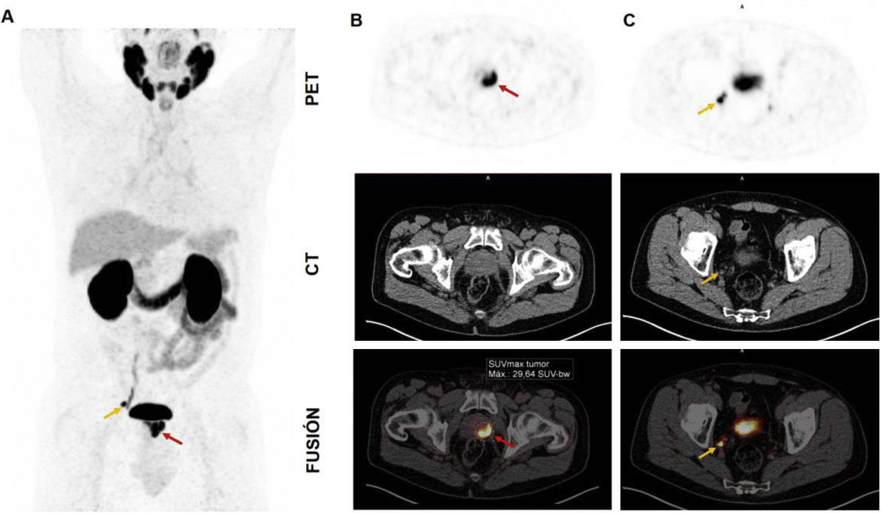
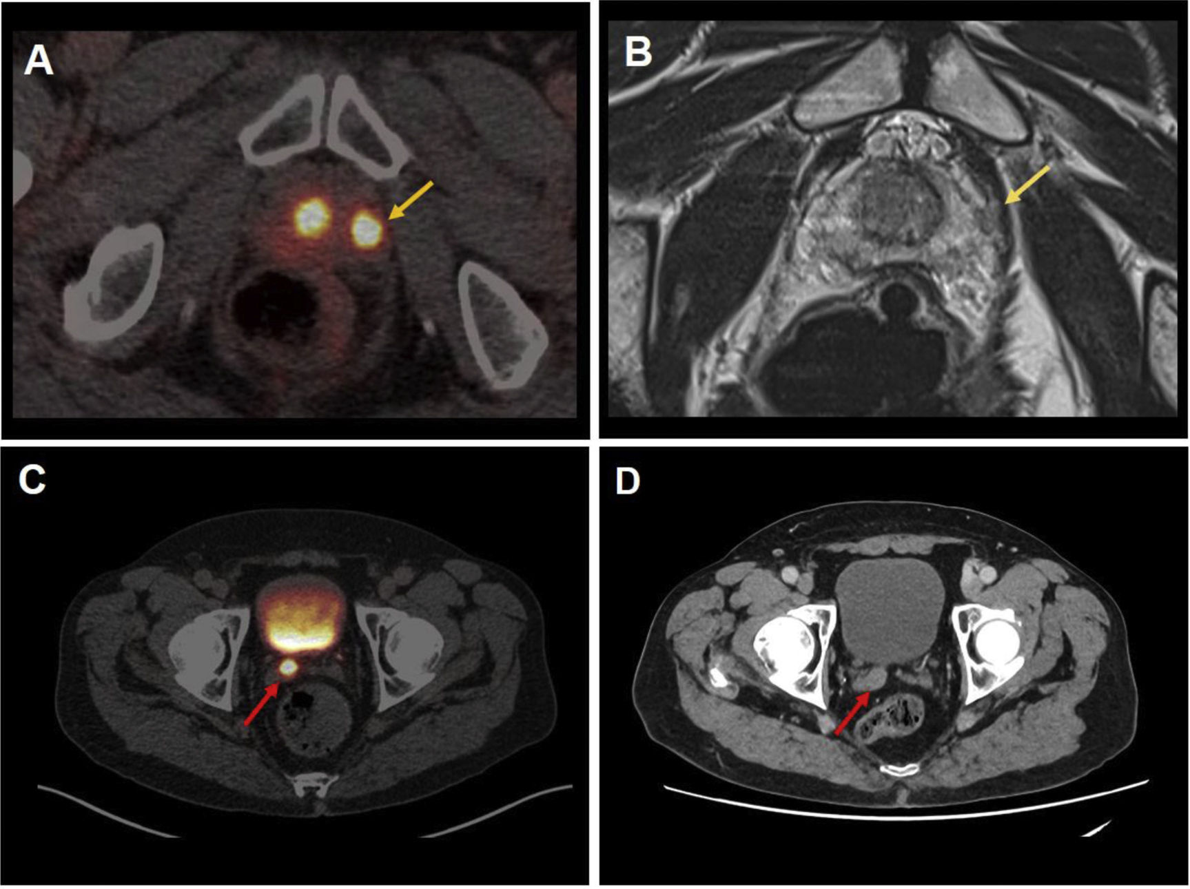
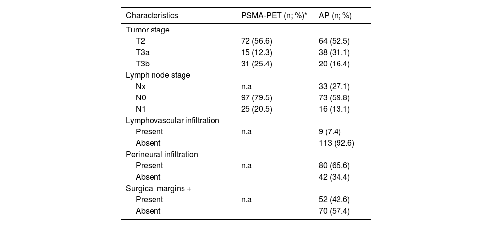
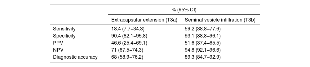

To evaluate the diagnostic accuracy of [68Ga]Ga-PSMA-11 PET/CT (PET-PSMA) in local and loco-regional nodal staging compared with histopathological results in intermediate- and high-risk prostate cancer patients treated with radical prostatectomy (RP) and pelvic lymph node dissection (PLND).
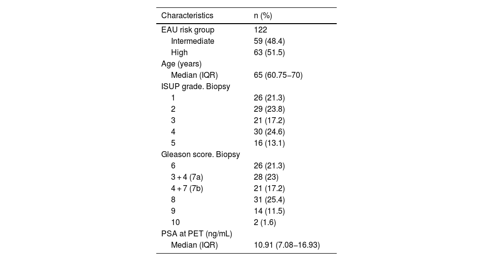
Materials y methodsA total of 122 intermediate- and high-risk prostate cancer (PCa) patients staged with PET-PSMA and treated with RP (36/122) and RP plus PLND (86/122) from December 2018 to December 2023 were included. Visual and semiquantitative analysis findings using the SUVmax of the molecular imaging were correlated with histopathological results.
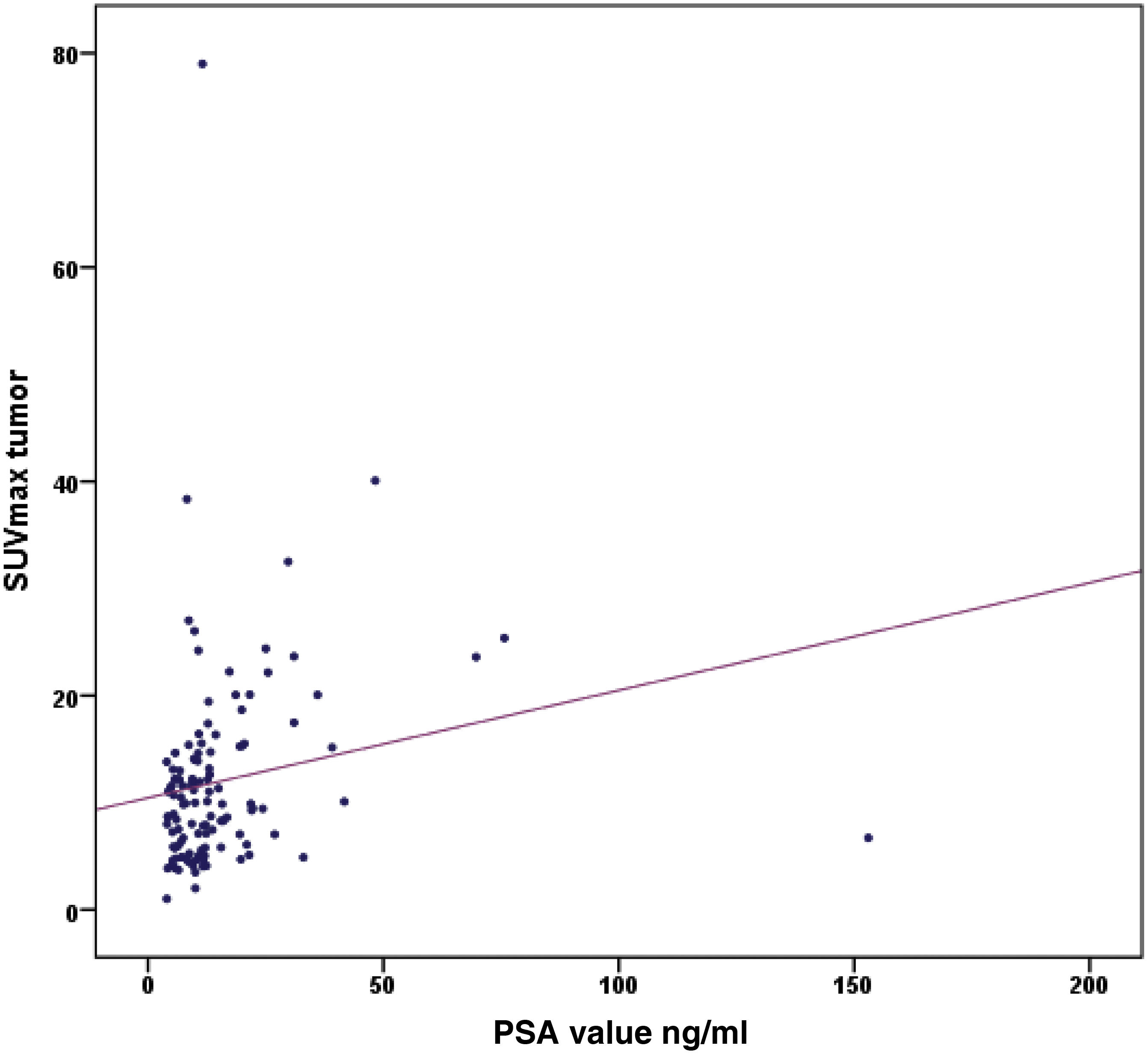
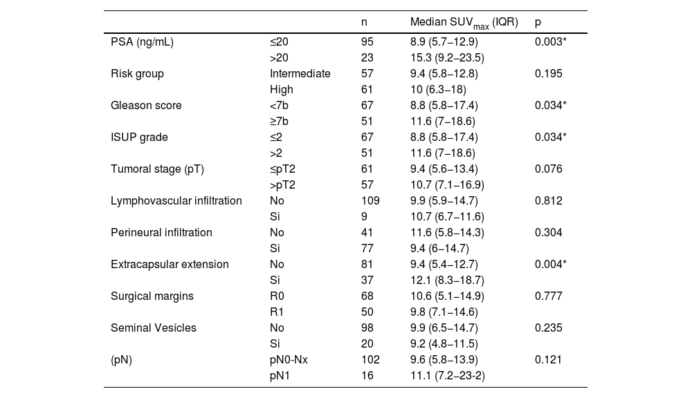
ResultsThe primary tumor was visible by PET-PSMA in 96.7% of the patients. A positive correlation was found between PSA levels and SUVmax (Spearman’s r: 0.303, p < 0.001). PET-PSMA detected nodal involvement in 25/89 patients (28.08%). The sensitivity, specificity, and diagnostic accuracy of PET-PSMA for detecting nodal involvement were 75%, 82.2%, and 80.9%, respectively. Patients with PSA levels >20 ng/mL, Gleason score ≥7b, ISUP grade >2, and extracapsular extension showed significantly higher SUVmax values. No differences were observed in SUVmax between risk groups or in other histopathological variables.
ConclusionsPET-PSMA is an effective tool for the initial staging of intermediate- and high-risk PCa. SUVmax values were significantly higher in patients with unfavorable clinical features.
Evaluar la precisión diagnóstica de la PET/TC con [68Ga]Ga-PSMA-11 (PET-PSMA) en la estadificación local y ganglionar loco-regional en comparación con los resultados anatomopatológicos en pacientes con cáncer de próstata (cPr) de riesgo intermedio y alto tratados con prostatectomía radical (PR) y linfadenectomía pélvica (LDNP).
Material y métodosSe incluyeron 122 pacientes con cPr de riesgo intermedio y alto estadificados mediante PET-PSMA y tratados mediante PR (36/122) y PR más LDNP (86/122) desde diciembre de 2018 hasta diciembre de 2023. Los hallazgos del análisis visual y semicuantitativo mediante el SUVmax de la imagen molecular se correlacionaron con los resultados anatomopatológicos.
ResultadosEl tumor primario fue visible mediante PET-PSMA en el 96,7% de los pacientes. Se encontró una correlación positiva entre los valores de PSA y SUVmax (r de Spearman: 0,303, p < 0,001). La PET-PSMA detectó infiltración ganglionar en 25/89 pacientes (28,08%). La sensibilidad, especificidad y precisión diagnóstica de la PET-PSMA para detectar infiltración ganglionar fue de 75%, 82,2% y 80,9% respectivamente. Los pacientes con valores de PSA > 20 ng/mL, grado de Gleason ≥7b, grado de la ISUP > 2 y extensión extracapsular mostraron valores de SUVmax significativamente mayores. No se observaron diferencias en el SUVmax por grupos de riesgo ni en el resto de variables anatomopatológicas.
ConclusionesLa PET-PSMA es una herramienta eficaz para la estadificación inicial del cPr de riesgo intermedio y alto. Los valores de SUVmax fueron significativamente mayores en pacientes con características clínicas desfavorables.
Article
If you experience access problems, you can contact the SEMNIM Technical Secretariat by email at secretaria.tecnica@semnim.es or by phone at +34 619 594 780.
Revista Española de Medicina Nuclear e Imagen Molecular (English Edition)