


To assess the diagnostic impact of using automatic segmentation and quantitative analysis of lung perfusion SPECT/CT in the evaluation of pulmonary reperfusion in patients undergoing follow-up for pulmonary thromboembolism (PTE).
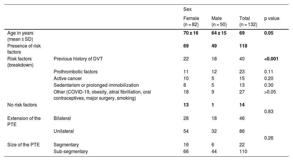
Materials and methodsA prospective study was conducted from October 2021 to October 2024. We included 132 patients with PTE diagnosed by lung perfusion scintigraphy with SPECT/CT, who underwent a follow-up scan at 6 months.
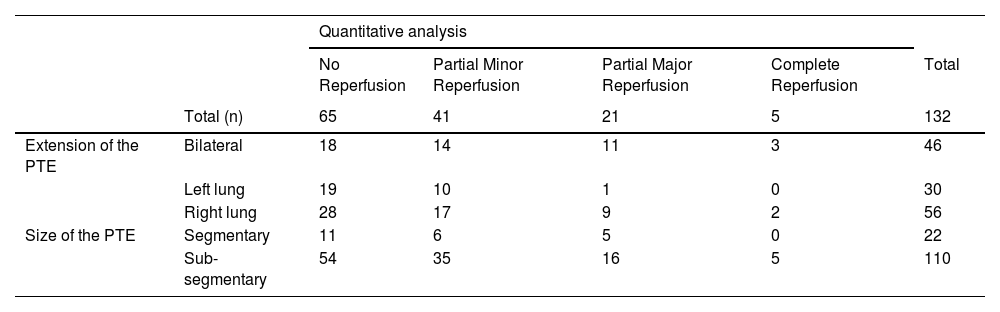
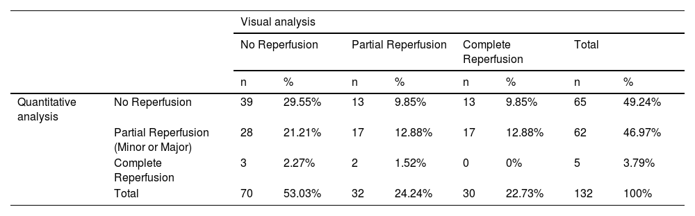
Reperfusion was assessed visually and quantitatively. Three grades were visually established: no reperfusion, partial reperfusion, and complete reperfusion. For quantitative analysis, automatic segmentation was performed, obtaining volumes and total counts in the baseline and follow-up SPECT/CT scans. Two parameters were established for comparison: the relative decrease in defect volume (RDV) and the relative increase in total perfused volume counts (RIC). Reperfusion was classified as: no reperfusion, partial reperfusion (minor and major), and complete reperfusion.
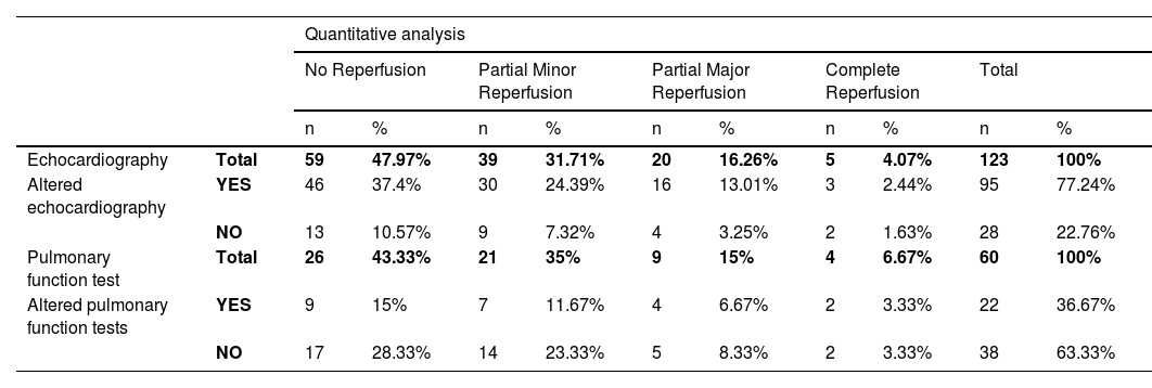
The scintigraphic results were correlated with demographic variables, extension and size of the PTE and other complementary diagnostic tools (pulmonary function tests and echocardiogram).
Results82 patients were women (mean age 70 ± 16 years) and 50 were men (64 ± 15 years).
Comparing the agreement between visual and quantitative analysis (weighted Cohen's Kappa index), a slight, but non-statistically significant, agreement was demonstrated between evaluators (κ = 0.04; p = 0.464).
Pearson's assessment revealed a very high and positive assessment between RDV and RIC (r = 0.77; p < 0.001).
Patients with abnormal complementary diagnostics tools at follow-up were not more likely to have residual thrombosis (p > 0.05 in the visual and quantitative analyses). The remaining variables also did not show statistical significance in the persistence of PTE.
ConclusionsIn the assessment of post-PTE pulmonary reperfusion, quantitative analysis of lung perfusion SPECT/CT is superior to visual analysis. Furthermore, it suggests that it is a particularly useful tool in patients in whom visual analysis does not show improvement, as it could prevent unnecessary and prolonged treatment if reperfusion is observed through quantification.
Valorar el impacto diagnóstico de utilizar la segmentación automática y el análisis cuantitativo del SPECT/TC de perfusión pulmonar en la evaluación de la reperfusión pulmonar de los pacientes en seguimiento por tromboembolismo pulmonar (TEP).
Material y métodosEstudio prospectivo, desde octubre 2021 hasta octubre 2024, en el que incluimos 132 pacientes con TEP diagnosticado por gammagrafía de perfusión pulmonar con SPECT/TC, a los que se les realizó una gammagrafía de seguimiento a los 6 meses.
La reperfusión se valoró de forma visual y cuantitativa. Visualmente se establecieron 3 grados: no reperfusión, reperfusión parcial y reperfusión completa. Para el análisis cuantitativo se realizó una segmentación automática obteniendo volúmenes y cuentas totales en el SPECT/TC basal y en el de seguimiento. Se establecen dos parámetros de comparación: la disminución relativa del volumen del defecto (DRV) y el aumento relativo de las cuentas totales del volumen perfundido (ARC). La reperfusión se clasificó en: no reperfusión, reperfusión parcial (menor y mayor) y reperfusión completa.
Se correlacionaron los resultados gammagráficos con variables demográficas, extensión y tamaño del TEP y otras pruebas complementarias (pruebas funcionales respiratorias y ecocardiograma).
Resultados82 eran mujeres (edad media 70 ± 16 años) y 50 hombres (64 ± 15 años).
Al comparar la concordancia entre el análisis visual y cuantitativo (índice Kappa de Cohen ponderado), se demostró una ligera concordancia entre los evaluadores no estadísticamente significativa (κ = 0,04; p = 0,464).
Mediante el análisis de correlación de Pearson se observó una correlación muy alta y positiva entre la DRV y el ARC (r = 0,77; p < 0,001).
Los pacientes que presentaban pruebas complementarias alteradas al seguimiento no tenían mayor probabilidad de trombosis residual (p > 0,05 en el análisis visual y cuantitativo). El resto de variables tampoco mostraron significación estadística en la persistencia del TEP.
ConclusionesEn la valoración de la reperfusión pulmonar pos-TEP, el análisis cuantitativo del SPECT/TC de perfusión pulmonar es superior al análisis visual. Además, sugiere ser una herramienta especialmente útil en los pacientes en los que no se objetiva mejoría en el análisis visual ya que podría evitar tratamientos innecesarios y prolongados si se observa reperfusión mediante la cuantificación.
Article
If you experience access problems, you can contact the SEMNIM Technical Secretariat by email at secretaria.tecnica@semnim.es or by phone at +34 619 594 780.

Revista Española de Medicina Nuclear e Imagen Molecular (English Edition)
