


18F-Fluoro-l-dihydroxyphenylalanine (18F-DOPA) PET offers high sensitivity and specificity in the imaging of non-malignant extra-adrenal paraganglioma (PGL) and pheochromocytoma (PHEO) but lower sensitivity in metastatic disease. These tumours are of neuroendocrine origin and can be detected by 68Ga-DOTA-Tyr3-octreotide (68Ga-DOTA-TOC) PET. Therefore, we compared 68Ga-DOTA-TOC and 18F-DOPA as radiolabels for PET/CT imaging for the diagnosis of metastatic extra-adrenal PGL and PHEO. Combined cross-sectional imaging was the reference standard.
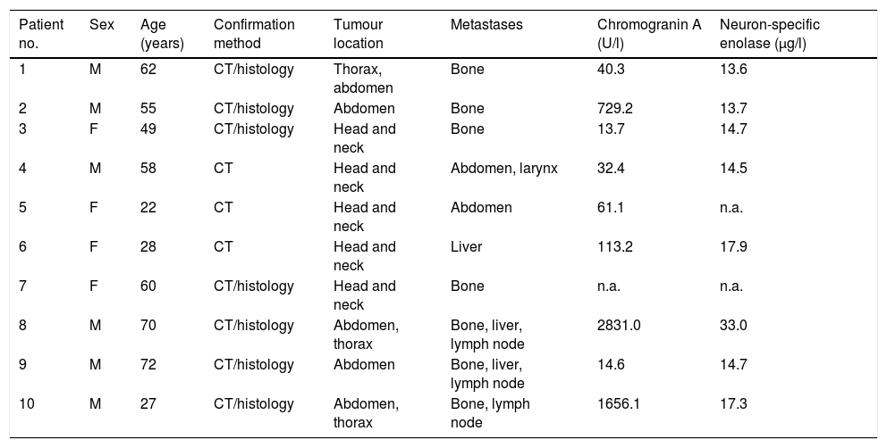
MethodsA total of 6 men and 4 women (age range 22–72 years) with anatomical and/or histologically proven metastatic PGL and PHEO were included in this study. Of these patients, 2 male patients suffered from PHEO, while the remaining 8 patients were diagnosed as metastatic extra-adrenal PGL disease. Comparative evaluation included morphological imaging with CT and functional imaging with 68Ga-DOTA-TOC and 18F-DOPA PET. The imaging results were analyzed on a per-lesion basis. The maximum standardized uptake value (SUVmax) of each functional imaging modality in concordant tumour lesions was measured.
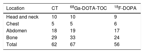
ResultsCompared with anatomical imaging, the per-lesion detection rate of 68Ga-DOTA-TOC was 100% (McNemar, P<0.01), and that of 18F-DOPA PET was 82.3% (McNemar, P<0.8) in metastatic extra-adrenal PGL and PHEO. Overall, 68Ga-DOTA-TOC PET identified 67 lesions; anatomical imaging identified 62 lesions, and 18F-DOPA PET identified 56 lesions. The SUVmax (mean±SD) of all concordant lesions was 29.3±19.9 for 68Ga-DOTA-TOC PET and 12.3±9.1 for 18F-DOPA PET (Mann–Whitney U test, P<0.0001).
Conclusion68Ga-DOTA-TOC PET offers the highest detection rate in metastatic extra-adrenal PGL and PHEO compared to 18F-DOPA PET and even to diagnostic CT, particularly in bone lesions. Combined functional/anatomical imaging (68Ga-DOTA-TOC PET/CT) enables exact tumour extension to be detected in these rare tumour entities, especially in the case of unclear anatomical correlation.
La PET con 18F-Fluoro-L-dihidroxifenilalanina (18F-DOPA) ofrece alta sensibilidad y especificidad en el diagnóstico del paraganglioma extra-adrenal no maligno (PGL) y el feocromocitoma (PHEO), pero menor sensibilidad en la enfermedad metastásica. Estos tumores son de origen neuroendocrino, y pueden detectarse mediante PET con 68Ga-DOTA-Tyr3-octreótida (68Ga-DOTA-TOC). Por tanto, comparamos 68Ga-DOTA-TOC y 18F-DOPA como radiotrazadores para PET/TC para el diagnóstico de PGL extra-adrenal metastásico y PHEO. Las imágenes tomográficas anatómicas y funcionales fusionadas se utilizaron como estándar de referencia.
MétodosSe incluyó en el estudio a un total de 6 varones y 4 mujeres (rango de edad de 22 a 72 años), con PGL metastásico y PHEO anatómica y/o histológicamente demostrados. De entre estos pacientes, dos varones padecían PHEO, y los ocho pacientes restantes PGL extra-adrenal metastásico. La evaluación comparativa incluyó imagen morfológica con TC, e imagen funcional mediante PET con 68Ga-DOTA-TOC y 18F-DOPA. Se analizaron los resultados de las imágenes por lesión. Se midió el valor máximo de captación estandarizado (SUVmax) de cada modalidad de imagen funcional en las lesiones tumorales concordantes.
ResultadosEn comparación con la imagen anatómica, la tasa de detección por lesión mediante PET con 68Ga-DOTA-TOC fue del 100% (McNemar, P<0,01), y la de PET con 18F-DOPA fue del 82,3% (McNemar, P<0,8) para PGL extra-adrenal metastásico y PHEO. En general, la PET con 68Ga-DOTA-TOC identificó 67 lesiones, la imagen anatómica identificó 62 lesiones, y la PET con 18F-DOPA identificó 56 lesiones. El valor SUVmax (media+/- DE) de todas las lesiones concordantes fue de 29,3+/- 19,9 para la PET con 68Ga-DOTA-TOC, y de 12,3+/- 9,1 para la PET con 18F-DOPA (prueba U de Mann-Whitney, P<0,0001).
ConclusiónLa PET con 68Ga-DOTA-TOC proporciona un índice de detección más elevado en el PGL extra-adrenal metastásico y PHEO, en comparación con la PET con 18F-DOPA e incluso con la TC diagnóstica, particularmente en lo referente a lesiones óseas. La imagen funcional/anatómica combinada (PET/TC con 68Ga-DOTA-TOC) permite detectar la extensión exacta del tumor en estas entidades tumorales infrecuentes, especialmente en caso de correlación anatómica incierta.
Article
If you experience access problems, you can contact the SEMNIM Technical Secretariat by email at secretaria.tecnica@semnim.es or by phone at +34 619 594 780.

Revista Española de Medicina Nuclear e Imagen Molecular (English Edition)
