


The aims of this study were to evaluate the relationships between textural features of the primary tumor on FDG PET images and clinical-histopathological parameters which are useful in predicting prognosis in newly diagnosed non-small cell lung cancer (NSCLC) patients.
MethodsPET/CT images of ninety (90) patients with NSCLC prior to surgery were analyzed retrospectively. All patients had resectable tumors. From the images we acquired data related to metabolism (SUVmax, MTV, TLG) and texture features of primary tumors. Histopathological tumor types and subgroups, degree of Ki-67 expression and necrosis rates of the primary tumor, mediastinal lymph node (MLN) status and nodal stages were recorded.
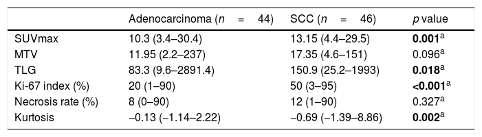
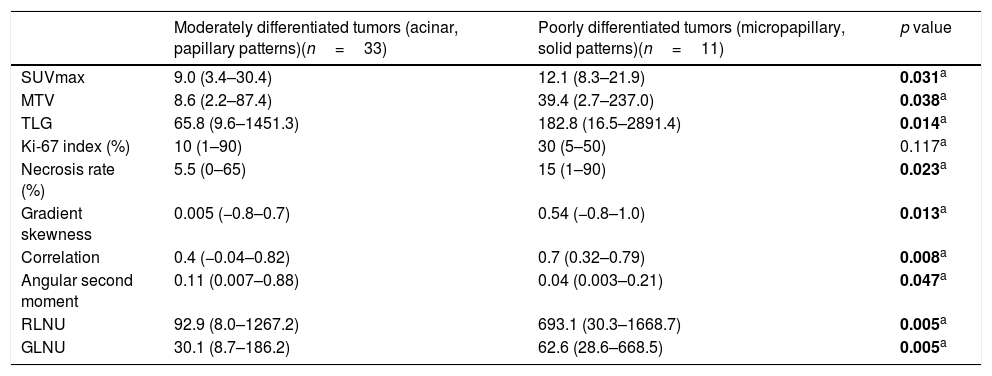
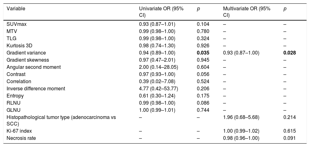
ResultsAmong the two histologic tumor types (adenocarcinoma and squamous cell carcinoma) significant differences were present regarding metabolic parameters, Ki-67 index with higher values and kurtosis with lower values in the latter group. Textural heterogeneity was found to be higher in poorly differentiated tumors compared to moderately differentiated tumors in patients with adenocarcinoma. While Ki-67 index had significant correlations with metabolic parameters and kurtosis, tumor necrosis rate was only significantly correlated with textural features. By univariate and multivariate analyses of the imaging and histopathological factors examined, only gradient variance was significant predictive factor for the presence of MLN metastasis.
ConclusionsTextural features had significant associations with histologic tumor types, degree of pathological differentiation, tumor proliferation and necrosis rates. Texture analysis has potential to differentiate tumor types and subtypes and to predict MLN metastasis in patients with NSCLC.
En este estudio evaluamos las relaciones entre las características texturales de los tumores primarios en las imágenes PET-FDG y los parámetros clínico-histopatológicos. Estos parámetros son útiles para predecir el pronóstico de nuevos pacientes diagnosticados con cáncer de pulmón de células no pequeñas (CPCNP).
Materiales y MétodosLas imágenes PET/TC de noventa (90) pacientes con CPCNP antes de la cirugía se analizaron retrospectivamente. Todos los pacientes tenían tumores resecables. De las imágenes adquirimos datos relacionados con el metabolismo (SUVmax, MTV, TLG) y las características de textura de los tumores primarios. Se registraron los tipos y subgrupos de tumores histopatológicos, el grado de expresión de Ki-67 y las tasas de necrosis del tumor primario, el estado de los ganglios linfáticos mediastínicos (MLN) y las etapas nodales.
ResultadosEntre los dos tipos de tumores histológicos (adenocarcinoma y carcinoma de células escamosas) hubo diferencias significativas con respecto a los parámetros metabólicos, el índice Ki-67 con valores más altos y la curtosis con valores más bajos en el último grupo. Se encontró que la heterogeneidad textural es mayor en los tumores poco diferenciados en comparación con los tumores moderadamente diferenciados en pacientes con adenocarcinoma. Mientras que el índice Ki-67 tenía correlaciones significativas con los parámetros metabólicos y la curtosis, la tasa de necrosis tumoral solo se correlacionó significativamente con las características de textura. Mediante análisis univariados y multivariados de los factores de imágen y los factores histopatológicos examinados, solo la variación del gradiente fue un factor predictivo significativo para la presencia de metástasis MLN.
ConclusiónLas características de textura tenían asociaciones significativas con los tipos de tumores histológicos, el grado de diferenciación patológica, la proliferación tumoral y las tasas de necrosis. El análisis de textura tiene el potencial de diferenciar los tipos y subtipos de tumores y predecir la metástasis de MLN en pacientes con CPCNP.
Article
If you experience access problems, you can contact the SEMNIM Technical Secretariat by email at secretaria.tecnica@semnim.es or by phone at +34 619 594 780.

Revista Española de Medicina Nuclear e Imagen Molecular (English Edition)
