


To review and classify the interval cancers found in the Principality of Asturias's Breast Cancer Screening Program (PDPCM). A secondary objective was to determine the histological characteristics, size, and stage of the interval cancers at the time of diagnosis.
Material and methodsWe included the interval cancers in the PDPCM in the period 2003–2007. Interval cancers were classified according to the breast cancer screening program protocol, with double reading without consensus, without blinding, with arbitration. Mammograms were interpreted by 10 radiologists in the PDPCM.
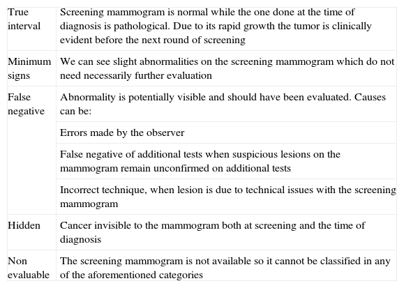
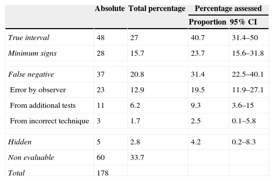
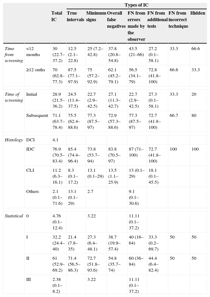
ResultsA total of 33.7% of the interval cancers could not be classified; of the interval cancers that could be classified, 40.67% were labeled true interval cancers, 31.4% were labeled false negatives on screening, 23.7% had minimal signs, and 4.23% were considered occult. A total of 70% of the interval cancers were diagnosed in the year of the period between screening examinations and 71.7% were diagnosed after subsequent screening. A total of 76.9% were invasive ductal carcinomas, 61.1% were stage II when detected, and 78.7% were larger than 10mm when detected.
ConclusionsThe rate of interval cancers and the rate of false negatives in the PDPCM are higher than those recommended in the European guidelines. Interval cancers are diagnosed later than the tumors detected at screening. Studying interval cancers provides significant training for the radiologists in the PDPCM.
Revisar y clasificar los carcinomas de intervalo (CI) del programa de detección precoz de cáncer de mama del Principado de Asturias (PDPCM). Como objetivo secundario se plantea la descripción de sus características anatomopatológicas, así como de su tamaño y estadio en el momento del diagnóstico.
Material y métodosSe incluyeron los CI del PDPCM correspondientes al período 2003-2007. Se clasificaron según el protocolo de los programas de detección precoz de cáncer de mama, mediante doble lectura sin consenso ni enmascaramiento, con arbitrio. Hubo 10 lectores diferentes, todos ellos radiólogos del PDPCM.
ResultadosNo pudo ser clasificado el 33,7% del total de CI; del resto, el 40,67% se etiquetaron de intervalos verdaderos, el 31,4% como falsos negativos, el 23,7% como signos mínimos y el 4,23% se consideraron ocultos. El 70% de los CI se diagnosticaron en el segundo año del período entre cribados y un 71,7% tras un cribado subsiguiente. El 76,9% resultaron carcinomas ductales infiltrantes, el 61,1% se detectó en estadio II, y el 78,7% eran mayores de 10mm cuando fueron diagnosticados.
ConclusionesLa tasa de CI y la proporción de falsos negativos son superiores a las recomendadas por las guías europeas de calidad. El diagnóstico del CI es más tardío que el de los tumores detectados dentro del PDPCM. El estudio de los CI conlleva una importante labor formativa para los radiólogos del PDPCM.

