


metricas
Cirugía Española (English Edition) Local resection in rectal cancer: When, who and how?
Journal Information
Visits
1043
Vol. 103. Issue 4.
Pages 179-254 (April 2025)
Special article
Full text access

Local resection in rectal cancer: When, who and how?

Resección local en cáncer de recto: ¿cuándo, a quién y cómo?
Visits
1043
Jesus Badia-Closaa, Juan Pablo Campanab, Gustavo Leandro Rossib, Xavier Serra-Aracilc,
Corresponding author
xserraa@gmail.com

Corresponding author.
a Unidad Colorrectal, Servicio de Cirugía General y Digestiva, Hospital de Sant Joan Despí Moisès Broggi, Barcelona, Spain
b Sección de Cirugía Colorrectal, Servicio de Cirugía General, Hospital Italiano de Buenos Aires, Buenos Aires, Argentina
c Unidad de Coloproctología, Hospital Universitario Parc Tauli, Sabadell. Institut d'investigació i innovació Parc Tauli I3PT-CERCA, Department of Surgery, Universitat Autònoma de Barcelona, Barcelona, Spain
Related content
This item has received
Article information
Abstract
Full Text
Bibliography
Download PDF
Statistics
Figures (4)
fig0005
fig0010
fig0015
fig0020
Tables (3)
Tables
Tables
Tables
Special issue
This article is part of special issue:
Rectal Cancer Treatment: The Future is already the Present

Edited by: Dr. Calos Cerdán
Cirugía General y Aparato Digestivo. Unidad de Cirugía Digestiva. Hospital Universitario de la Princesa. Madrid
Dr. Matteo Frasson
Cirugía general. Hospital Universitario y Politécnico La Fe. Valencia

Last update: January 2026

More info
Abstract

Local resection (LR) in rectal cancer is indicated in stage T1N0M0 without unfavorable pathological factors, achieving oncologically satisfactory outcomes through transanal endoscopic surgery techniques. However, the initial step involves accurate staging and selection of these tumors through specific tests conducted in specialized colorectal units.

For T2N0M0 tumors and T1 tumors with poor prognostic factors, the standard treatment is total mesorectal excision (TME), a procedure associated with high postoperative morbidity and mortality, functional impairments, and reduced quality of life. Therefore, new organ-preservation strategies are being explored as alternatives to TME. These include neoadjuvant therapy combined with LR, which has shown promising results, and neoadjuvant therapy followed by a “Watch and Wait” approach –where patients with complete clinical response are selected for strict surveillance– as an ideal future treatment, although there are still current challenges to be addressed.

Keywords:
Rectal cancer
Early rectal cancer
Local resection
Transanal endoscopic surgery
Total mesorectal excision
Neoadjuvant therapy and rectal cancer
Resumen

La resección local (RL) en el cáncer de recto está indicada en el estadio T1N0M0 sin factores de riesgo anatomopatológicos, que con técnicas de cirugía endoscópica transanal obtiene resultados oncológicamente satisfactorios. Sin embargo, el primer paso es una correcta estadificación y selección de estos tumores mediante pruebas específicas realizadas en unidades especializadas en colorrectales.

En los tumores T2N0M0 y T1 con factores de mal pronóstico, el tratamiento estándar es la escisión total del mesorrecto (ETM), procedimiento que se asocia a una elevada morbi-mortalidad postoperatoria, alteraciones funcionales y de calidad de vida. Por ello, nuevas estrategias de preservación de recto se encuentran actualmente como alternativa a la ETM. Entre ellas se incluyen la neoadyuvancia asociada a la RL con resultados esperanzadores y la neoadyuvancia seguida de la selección de pacientes con respuesta clínica completa y seguimiento estricto, denominada "Watch and Wait”, como tratamiento futuro idóneo pero con cuestiones actuales a resolver.


Palabras clave:
Cáncer de recto
Cáncer de recto precoz
Resección local
Cirugía endoscópica transanal
Excision total del mesorrecto
Neoadyuvancia y cáncer de recto
Full Text
Introduction

Although total mesorectal excision (TME) remains the standard of care for most patients with rectal cancer, the increasing incidence of early rectal cancer has led to increased interest in local excision and organ preservation techniques for the treatment of these patients.1 Transanal local resection (LR) has important advantages in terms of postoperative morbidity and quality of life,2,3 which is what makes it attractive.

In recent years, the advent of transanal devices such as TAMIS (TransAnal Minimally Invasive Surgery), TEM (Transanal Endoscopic Microsurgery), and TEO (Transanal Endoscopic Operation) has radically improved the quality of resection, allowing specimens that are complete, larger and further from the anal margin.4 This has improved the oncological outcomes of this technique, bringing them closer to those of much more radical surgery.5

While LR can achieve resection of the primary tumour with adequate margins, the lack of associated lymphadenectomy has limited its use to patients with superficial tumours and no evidence of lymph node metastasis. Indeed, historical series have shown discouraging results in some pT1 and in pT2.6,7 However, with the introduction of new neoadjuvant chemoradiotherapy (CRT) strategies, the indications for LR have expanded to include patients initially not considered candidates for LR·8

This article aims to provide a comprehensive review of the historical evolution, technical rationale, clinical outcomes, and future implications of LR in the treatment of rectal tumours as an integral part of organ preservation strategies.

Diagnosis of rectal lesions

Accurate diagnosis of rectal lesions is essential to determine the most appropriate treatment and to predict disease progression. The main limitation of LR is that only the endoluminal tumour is resected, leaving the mesorectum intact with its lymph node component, which may increase the risk of local and distant recurrence. For this reason, it is essential to identify risk factors for lymph node metastasis during preoperative staging in order to properly select patients who will truly benefit from this approach.

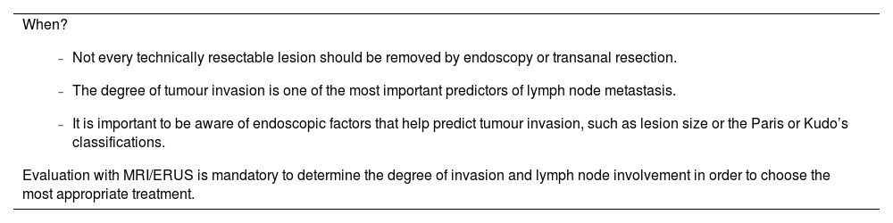
The degree of tumour invasion is one of the most important predictors of lymph node metastasis. If we divide submucosal invasion into thirds (Kikuchi classification), the risk varies significantly, at 0%–3% in pT1 sm1 (superficial third), 8%–11% in sm2 (superficial two thirds), and 10%–23% in sm39,10 (deep third involvement). The risk of lymph node involvement in pT2 tumours was close to 20%,11 suggesting that pT1 sm2 and sm3 tumours behave more like pT2 tumours than pT1 tumours. Other important features associated with the risk of lymph node involvement are the degree of tumour differentiation, lymphovascular invasion, and more distal location in the rectum.12

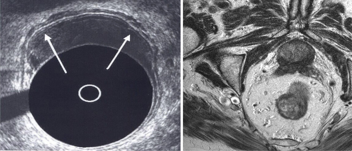
Diagnosis of distant disease is initially made by computed tomography. Local assessment of the tumour requires digital examination in conjunction with imaging techniques. Three imaging modalities are widely used in this context: endoscopy, magnetic resonance imaging (MRI), and endorectal ultrasound (ERUS) (Fig. 1). Each of these techniques offers significant advantages in staging rectal lesions and assessing the local extent of the tumour.

Fig. 1.

Endorectal ultrasound and MRI image of a cT1 tumour.

In early rectal cancer, endoscopic evaluation is essential for the final treatment decision. Firstly, to exclude synchronous lesions, which may be present in 3% of cases.13 Secondly, endoscopic classification of the lesion using the Paris, Kudo, or NICE scales can predict the possibility of submucosal invasion with high sensitivity and specificity,14,15 These classifications are becoming increasingly important as endoscopy is the first diagnostic test in many cases and endoscopic resection techniques have greatly improved in recent years, leading to a higher rate of lesion resection. Therefore, predicting those rectal lesions with a higher likelihood of invasion is crucial to avoid inadequate treatment that may later condition surgical resection. According to the Paris classification, superficial colorectal neoplasms can be classified as polypoid (sessile (Is) or pedunculated (Ip), or non-polypoid (flat raised (IIa), flat (IIb), and flat depressed (IIc)). The larger the size of the polypoid lesion, the greater the risk of deep invasion. In contrast, in non-polypoid lesions, size has less of an effect on the risk of deep invasion (in type IIc lesions, there is a risk of invasion even in lesions <1 cm).

Kudo’s pit pattern classification is based on the pattern of mucosal crypts and is divided into: non-neoplastic (type I or II, not requiring treatment), non-invasive neoplastic (type IIIS, IIIL, IV or selected cases of VI, corresponding to adenomas and cancers with superficial submucosal invasion, suitable for endoscopic treatment) and invasive (type VN and some VI, requiring surgery due to deep submucosal invasion).

Non-contrast diffusion-weighted MRI of the rectum is the method of choice for local staging of rectal tumours, especially in intermediate or locally advanced disease, as differentiation between submucosal invasion and muscularis propria is difficult.16 MRI is able to assess all pelvic lymph node compartments with a wider field of view than ERUS (Fig. 1). In addition to size, MRI allows assessment of morphological features of the lymph nodes, which may predict lymph node involvement,17 and assessment of vascular structures to determine the presence of extramural vascular invasion. The diffusion sequence improves nodal detection and facilitates response assessment after neoadjuvant treatment.

ERUS offers higher resolution than MRI, particularly in assessing the depth of invasion into the rectal wall. Some studies have confirmed these advantages,16,18 although high inter-operator variability and a long learning curve limit its use.19,20

When?
  • -

    Not every technically resectable lesion should be removed by endoscopy or transanal resection.

  • -

    The degree of tumour invasion is one of the most important predictors of lymph node metastasis.

  • -

    It is important to be aware of endoscopic factors that help predict tumour invasion, such as lesion size or the Paris or Kudo’s classifications.

Evaluation with MRI/ERUS is mandatory to determine the degree of invasion and lymph node involvement in order to choose the most appropriate treatment. 

Staging of rectal cancer

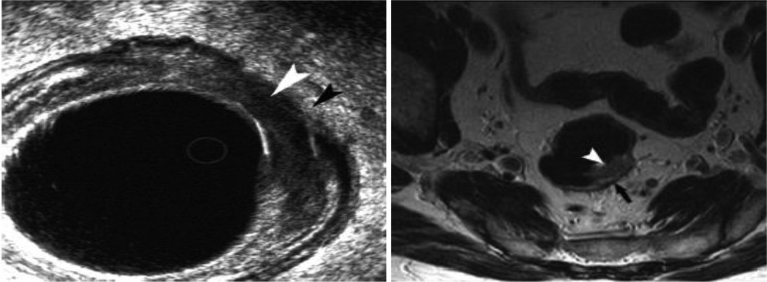
Stage I rectal cancer is the earliest stage and includes those adenocarcinomas that invade the muscularis mucosae of the rectal wall but are confined to the submucosa (T1) or muscularis propria (T2), without lymph node or distant involvement (N0, M0) (Fig. 2). Stage II defines rectal tumours that extend beyond the muscularis propria into the serosa or pericolic or perirectal tissues, without peritoneal involvement (T3) or with invasion into surrounding organs or tissues through the visceral peritoneum (T4), without lymph node or distant involvement (N0, M0). Stage III defines tumours with lymph node metastases, regardless of the depth of invasion. Finally, stage IV includes tumours with distant metastases.16,21

Fig. 2.

Endorectal ultrasound and MRI image of a cT2 tumour. The arrows show the muscularis propia.

The usefulness of staging lies in its correlation with five-year survival.22 Patients with stage I disease have excellent survival rates, >90% in most series. Survival decreases progressively with increasing stage, reaching approximately 15%–30% at five years in stage IV patients.

Rectal tumours amenable to LR, which are the focus of this review, include T1, T2 tumours without lymph node or distant involvement. These tumours infiltrate up to the muscularis propria, as described above. LR with complete wall excision can remove the entire tumour, leaving safe margins, so a priori this resection could be curative. However, as explained in the previous section, the potential for lymph node involvement, despite the fact that it may not be apparent on initial examination, limits the exclusive use of this technique in T2 tumours (around 20% N+) and in T1 tumours with poor prognostic factors. The proposed management and current evidence for these tumours is discussed in more detail below.

Treatment strategies for very early T1N0M0 tumours

LR for pT1 tumours without other associated risk factors has been shown to have similar long-term oncological outcomes to TME, but with less surgical morbidity.23 However, when the predominant selection criteria are fitness and comorbidities, without consideration of microscopic risk factors associated with the lesion, oncological outcomes are not favourable.24 In a retrospective study comparing radical surgery with LR for T1 tumours, local recurrence was significantly higher in the former group (13.2% vs. 2.7%, p = .001), with a lower cancer-related 5-year survival rate (87% vs. 96%, p = .03).6 Other studies have confirmed these findings, even suggesting that the risk of local recurrence may be more than 6-fold in this group.25 This suggests that those T1 lesions with unfavourable histological and morphological features may not be suitable for LR. However, the interpretation of these results must be balanced against the potential postoperative complications and functional changes associated with radical treatment. In a systematic review, LR was associated with higher local recurrence but lower perioperative mortality (RR .31, 95% CI .14–.71), fewer major postoperative complications (RR .20, 95% CI .10–.41) and less need for a permanent stoma (RR .17, 95% CI .09–.30) than radical surgery.26

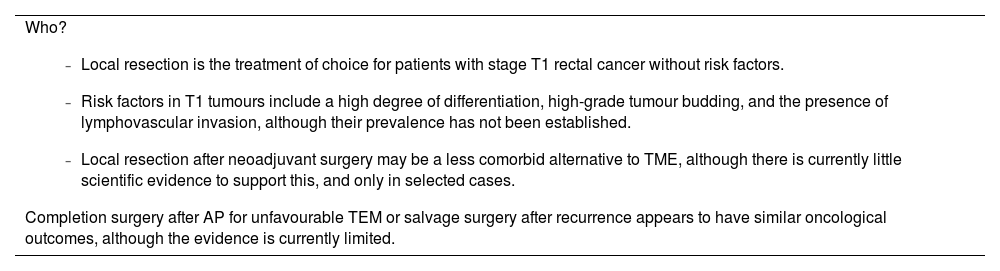
The indication for LR as the sole treatment for early rectal cancer depends primarily on the risk of lymph node metastasis from the lesion. The main risk factors identified are depth of tumour invasion, high differentiation, presence of lymphovascular invasion, and tumour budding.27 LR as a curative treatment for rectal cancer (T1) without these risk factors has been associated with high cancer-specific survival rates, which would justify its indication (Fig. 1). In contrast, the high rate of local recurrence in patients with rectal cancer associated with high risk factors limits the use of LR as the sole therapeutic measure.27

Recently, the true role of tumour invasion as an independent risk factor for recurrence has been reconsidered. In a systematic review including 67 studies (21,238 patients), submucosal invasion was not shown to be a significant independent predictor of nodal spread (OR, 1.73; 95% CI, .96−3. 12), in contrast to high-grade differentiation (OR, 2.14; 95% CI, 1.39–3.28), high-grade budding (OR, 2.83; 95% CI, 2.06–3.88), and the presence of lymphovascular invasion (OR, 3.16; 95% CI, 1.88–5.33).28 As a limitation of this systematic review, the authors acknowledge the wide variability of the included studies in terms of anatomical pathological analyses and the low quality of the reviewed articles. In addition, there is significant variability in the measurement of submucosal invasion due to the absence of muscularis mucosae in most cases. This supports the proposal to measure invasion from the muscularis propria in full thickness resection specimens.29

Sometimes, the preoperative histological diagnosis does not allow cT1 tumours to be classified as low or high risk depending on the presence or absence of the abovementioned risk factors. Clinical trials, such as the LORENA trial,30 are currently underway to determine the true prevalence of these poor prognostic factors in order to better predict the prognosis of these patients preoperatively and the potential need for TME. Because of this situation, some authors recommend the use of LR as an excisional biopsy of the lesion to obtain a more complete surgical specimen for histological evaluation. However, the use of LR as a diagnostic method may conflict with the need to complete surgery with TME if the histological findings are unfavourable.

Firstly, it should be considered that after full thickness LR, the scarring of the dissection bed may anatomically alter the surgical plane of the mesorectal fascia in the event of potential need for subsequent surgery, mainly on the anterior aspect of the rectum. Some authors have postulated that this makes the procedure more difficult, associated with higher morbidity and lower quality of the surgical specimen.31 Therefore, LR should not include perirectal fat. To counter this possible situation, the strategy of LR following the intermuscular plane has been postulated, avoiding entering the mesorectum and consequently altering this anatomy.32 Secondly, it has been observed that in patients with tumours very close to the pectineal line resected by LR and who subsequently need completion surgery with TME, the chances of sphincter preservation decrease.33 However, in referral centres, completion surgery showed similar results to primary TME in terms of major morbidity, abdominoperineal resection rate, and 3-year oncological outcomes.34 Therefore, LR as a diagnostic tool followed by completion surgery for early rectal cancer could be considered as a treatment option for rectal cancer in specialist centres for selected cases.

Treatment strategies for stage T2N0M0

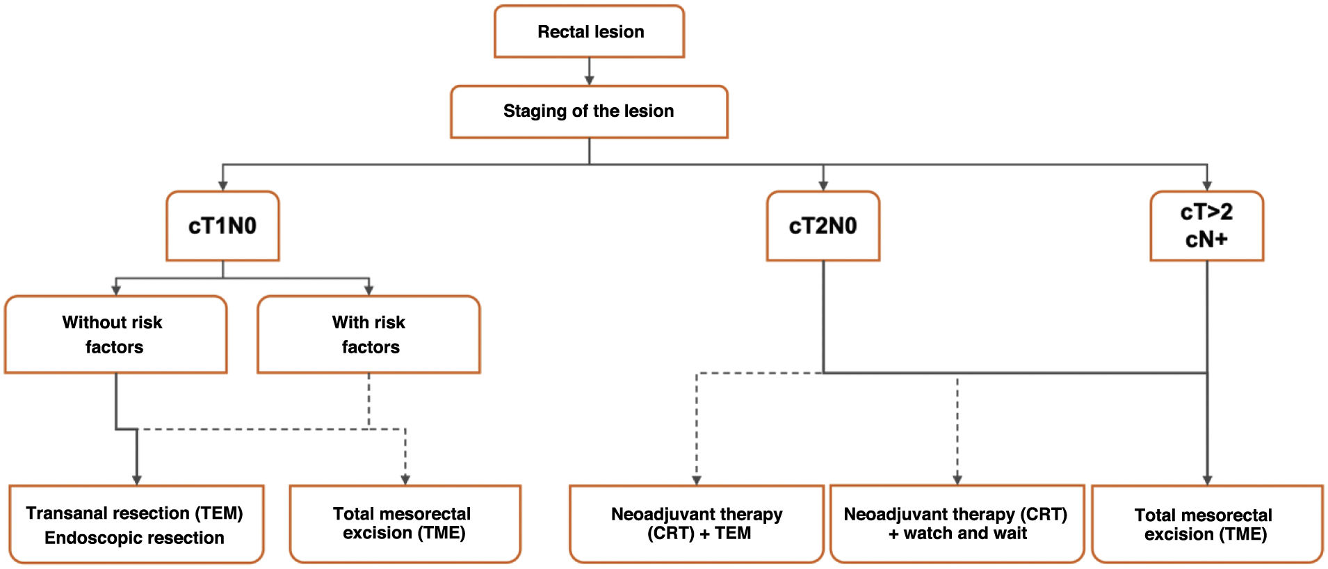
The possible strategies (surgical and medical) are described, indicating which have been shown to be most effective according to stage, including new strategies that have recently emerged and have changed the treatment strategy for rectal cancer at this stage. Fig. 3 shows the proposed treatment algorithm based on current evidence.

Fig. 3.

Proposed treatment algorithm in rectal cancer.

TME (Gold standard)

The therapeutic goal in rectal cancer is to remove the primary tumour together with its lymphovascular supply (including in the mesorectum) and any other surrounding organs or tissues invaded by the tumour. As such, TME is the conventional treatment for rectal cancer that manages to control locoregional disease, with a local recurrence rate of around 5% at 2 years.35

Despite its good oncological results, it is a technique associated with significant morbidity, which can occur in 30%–40%, with a non-negligible mortality of around 2%. The most common long-term complications include genitourinary changes and sexual dysfunction, as well as digestive changes, which are included in the syndrome known as LARS (Low Anterior Resection Syndrome).36

Depending on the height of the rectum where the tumour is located, the surgical technique varies. From high anterior resection (HAR) of the rectum with partial excision of the mesorectum for high rectal tumours, to low anterior resection (LAR), intersphincteric resection, transanal resection (TaTME), and abdominoperineal resection for tumours in the middle and lower third.

All these techniques can be performed using open, laparoscopic, laparoscopic combined with transanal and, more recently, robotic approaches.37,38

LR + adjuvant CRT: poor outcomes

As explained above, the treatment for early-stage disease (T1, without poor prognostic factors) is LR. As it is a less invasive treatment and therefore associated with less morbidity than TME, it has also been tried in more advanced stages. In stage T2-T3a-b, this treatment has a local recurrence rate of more than 20%, which is too high to be considered a valid treatment in these cases. This is why it has been postulated that a strategy could include the combination of adjuvant treatment (postoperative CRT) when the anatomo-pathological outcome after LR is T1 with poor prognostic factors or T2, thus avoiding TME. However, the oncological outcomes associated with this strategy have not been as promising as initially expected. In a systematic review published by Borstlap et al.,39 local recurrence rates in patients treated with this strategy were significantly higher than those treated with the gold standard (TME), almost twice as high (14% vs. 7%), no difference being found for distant recurrence (9% in both groups). For this reason, this strategy is no longer recommended from an oncological point of view and is almost exclusively reserved for elderly patients who have exceeded their life expectancy and for whom the oncological issue may become less important than safety and quality of life.

Neoadjuvant CRT + LR: Lines of investigation (TAU-TEM)

CRT treatment prior to TME has been shown to reduce local recurrence and increase survival in patients with locally advanced rectal cancer.40 This treatment can achieve a partial response in approximately 54%–75% of patients and a pathological complete response in 8%–27%.41 The absence of tumour after CRT has led to a growing interest in alternative rectal sparing strategies that avoid the adverse effects of TME.

Although there are several single-centre, observational, and retrospective studies in the literature with favourable outcomes for rectal preservation in stage T2N0M0,42 there are few prospective multicentre studies on this topic. All of them have different designs with different inclusion criteria and CRT regimens, achieving favourable but inconclusive outcomes.43,44,45,46

The TAU-TEM study sponsored by Serra-Aracil et al.,36,47 aimed to demonstrate non-inferiority of treatment with CRT and LR versus standard treatment with TME. The postoperative and anatomopathological results were presented in 2023. Organ preservation was achieved in 82.7% of patients in the experimental group, with a pathological complete response (pCR) rate of 44.3%. Quality of life (QoL) analysis in these patients using standardised tests (EORTC QLQ-C30, EORTC QLQ-CR38, and Karnofsky) showed a better QoL in patients who underwent CRT-LR compared to patients who underwent TME. Definitive oncological outcomes (analysis of local and systemic recurrence) will be published in 2025 and will show whether treatment with CRT + LR is a viable alternative to TME.

Other clinical trials such as ACOSOG Z6041 by Garcia-Aguilar et al. or GRECCAR 2 sponsored by Rullier et al. have also suggested that neoadjuvant treatment followed by LR may be an alternative to TME.

ACOSOG Z6041 is a phase II study in patients with small T2N0 tumours (≤4 cm in diameter) in the distal rectum. The results showed that most of these patients treated with neoadjuvant chemoradiotherapy and LR can preserve the rectum and achieve a survival equivalent to that of patients treated with TME, with minimal impact on anorectal function and quality of life. Similarly, GRECCAR 2 compared LR with TME in small (≤ 4 cm) T2/T3 N0-1 tumours of the distal rectum that respond well to neoadjuvant treatment. The pathological complete response rate in these tumours was 40%, demonstrating that they may be good candidates for organ preservation. The post-neoadjuvant LR strategy was oncologically safe, as 3-year local recurrence and disease-free survival did not differ between groups. Although the protocol required many patients undergoing LR to complete total mesorectal excision, the finding of positive mesorectal nodes was 8%, suggesting that completion surgery should be limited to less than 10% of patients (at stages ypT2/N1 and ypT3).

Neoadjuvant CRT + watch-and-wait: current evidence. Tests to be performed during follow-up

In recent years, a strategy called watch-and-wait (W&W), proposed by Habr-Gama et al. in 2004 has been promoted.48 This strategy is based on the response of some patients to neoadjuvant treatment. The clinical (rectoscopy, colonoscopy) and radiological (MRI) disappearance of the tumour is called clinical complete response (cCR) and is associated with favourable oncological outcomes. This strategy consists of identifying patients with a cCR and closely following them clinically without surgical intervention.

However, the correct preoperative identification of cCR remains a challenge, as it does not always coincide with pathological complete response (pCR49,50,51 (Fig. 2). Especially when the local regrowth rate in W&W has been described to be around 20% in T2,52,53 albeit with satisfactory overall and disease-free survival outcomes.54

Intensive follow-up of these patients is the key to early diagnosis of local recurrence. This makes the diagnostic tests to be performed particularly important. Current evidence suggests that follow-up should be based on a tri-modal approach: clinical, radiological, and endoscopic. Clinical follow-up consists of a clinical examination, which should include a digital rectal examination to confirm suspicion of regrowth.55 These techniques are far from infallible in assessing these lesions, as MRI has a sensitivity of around 90%, which decreases significantly to around 70% after administration of neoadjuvant treatment.47

Completion surgery or salvage surgery. Definition and differences

After LR, it is important to know the anatomopathological outcome as this will determine the need for further treatment. In this sense, it is important to prepare structured anatomopathological reports for this type of surgical specimen (including the most important aspects in terms of involvement and distance of margins, depth of invasion, etc.), as they facilitate the interpretation of the results and the homogeneity and comparability between different centres or studies. In cases where analysis of the resection specimen shows a more advanced stage than indicated by preoperative tests, or poor prognostic features (such as involved margins, angiolymphatic, or perineural invasion, poorly differentiated tumours), radical surgical treatment by TME is recommended. This adjuvant surgery has been termed completion surgery.56 Although results from prospective, randomised trials are not available, current evidence suggests that these patients have similar morbidity and survival to those who initially receive radical surgical treatment.33 Local resection appears to increase the risk of mesorectal scarring, which increases subsequent surgical difficulty and postoperative morbidity; therefore, resection of planned submucosal dissections has been suggested to avoid such scarring. Some studies suggest that patients who undergo LR followed by completion surgery are at higher risk of definitive colostomy compared with patients who undergo TME straight away.33,57 There is also insufficient evidence at present to know what is the appropriate treatment in case of recurrence or regrowth during follow-up in W&W, as some authors propose LR as a feasible alternative to classical TME.

As we mention above, strategies to preserve the rectum (CRT + TEM or W&W) have been a great advance and seem to be the future in the treatment of rectal cancer, but at the moment they are not free of potential complications. The real impact of performing completion surgery versus surgery when recurrence has already occurred (salvage surgery) remains to be determined.56

The local recurrence rate after completion surgery is around 5%, and similar after salvage surgery at 3%.56 Systemic recurrences have been found to be increased in patients with regrowth during follow-up compared with those without recurrence (18% vs. 5% according to the International Watch and Wait Database),58 although it is still not known whether this increase is due to the fact that patients with an incomplete response after CRT have an increased risk of metastatic disease, or whether the fact that TME was not performed early may also lead to an increased risk of metastatic disease

New, well-designed trials that focus on new neoadjuvant guidelines that achieve a higher percentage of pCR and evidence confirming the concordance between cCR and pCR will shape the future of rectal cancer treatment. This will allow organ preservation, less therapeutic aggression, and better quality of life for patients.

Who?
  • -

    Local resection is the treatment of choice for patients with stage T1 rectal cancer without risk factors.

  • -

    Risk factors in T1 tumours include a high degree of differentiation, high-grade tumour budding, and the presence of lymphovascular invasion, although their prevalence has not been established.

  • -

    Local resection after neoadjuvant surgery may be a less comorbid alternative to TME, although there is currently little scientific evidence to support this, and only in selected cases.

Completion surgery after AP for unfavourable TEM or salvage surgery after recurrence appears to have similar oncological outcomes, although the evidence is currently limited. 

LR techniques: Open resection, TEM, TEO, and TAMIS

As described above, LR can be performed using different approaches. After the open or "classic" approach, in which LR is performed using an anal retractor to improve visualisation of the lesions, devices have emerged that allow resection of a greater number of tumours, especially those that are further from the anal margin and where open resection has clear limitations.

The TEM and TEO platforms (practically identical, although the latter uses instruments compatible with conventional laparoscopic towers, which reduces its cost) use a rigid metal device between 7.5 and 20 cm in length. These devices provide a static view as they allow the optics to be fixed, facilitating the resection of lesions at a greater distance from the anal canal, introducing or removing the resectoscope as required. The instruments are usually used with their own instruments, with an angulation at the tip to facilitate manoeuvrability inside the rectum.

The TAMIS platform uses equipment available in conventional laparoscopic surgery, which dramatically reduces the cost of this platform. The GelPOINT® port, which is 4−5 cm long and allows the introduction of conventional laparoscopic instruments, is typically used. The shorter length allows for better manoeuvrability within the rectum, but limits the maximum distance of lesions that can be resected with this approach.

The choice of one platform or the other is based on the surgeon's experience and the availability of the platform, as there are no differences between them.

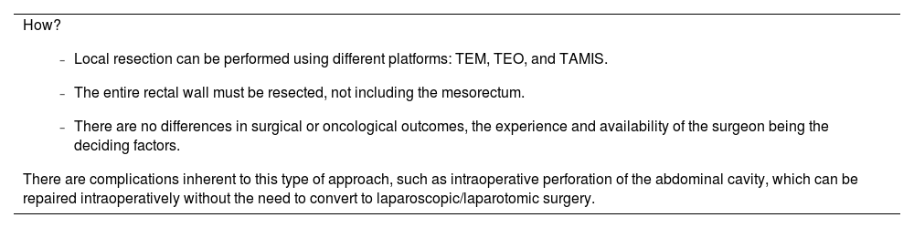
There can be complications from this approach, such as rectal perforation into the peritoneal cavity or intraluminal bleeding, which can be treated using the same approach without the need to convert to laparoscopy or laparotomy.59

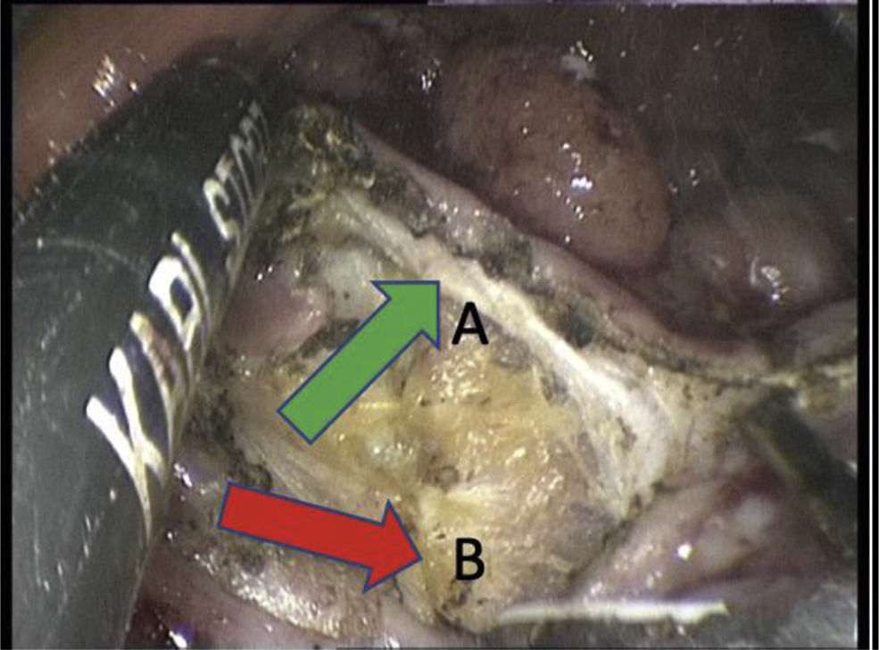
It is very important to emphasise the way complete wall resection is performed. This should be limited to resection of the rectal wall, without entering the perirectal fat, in order to preserve the integrity of the mesorectum in case it is necessary to complete the surgery with an TME (Fig. 4).

How?
  • -

    Local resection can be performed using different platforms: TEM, TEO, and TAMIS.

  • -

    The entire rectal wall must be resected, not including the mesorectum.

  • -

    There are no differences in surgical or oncological outcomes, the experience and availability of the surgeon being the deciding factors.

There are complications inherent to this type of approach, such as intraoperative perforation of the abdominal cavity, which can be repaired intraoperatively without the need to convert to laparoscopic/laparotomic surgery. 

Fig. 4.

Complete wall resection. The ultrasonic scalpel is used to cut the rectal wall down to the perirectal fat. This continues in this plane just below the muscular layer (arrow A) without entering the perirectal fat (arrow B).

References
[1]
V. Tonini, M. Zanni.
Why is early detection of colon cancer still not possible in 2023?.
World J Gastroenterol., 30 (2024), pp. 211-224
[2]
E. Lezoche, A.M. Paganini, B. Fabiani, A. Balla, A. Vestri, L. Pescatori, et al.
Quality-of-life impairment after endoluminal locoregional resection and laparoscopic total mesorectal excision.
Surg Endosc., 28 (2014), pp. 227-234
[3]
M.R. Albert, S.B. Atallah, T.C. deBeche-Adams, S. Izfar, S.W. Larach.
Transanal minimally invasive surgery (TAMIS) for local excision of benign neoplasms and early-stage rectal cancer: efficacy and outcomes in the first 50 patients.
Dis Colon Rectum, 56 (2013), pp. 301-307
[4]
C. Clancy, J.P. Burke, M.R. Albert, P.R. O’Connell, D.C. Winter.
Transanal endoscopic microsurgery versus standard transanal excision for the removal of rectal neoplasms: a systematic review and meta-analysis.
Dis Colon Rectum., 58 (2015), pp. 254-261
[5]
W. Li, X.X. Xiang, H. Da Wang, C.J. Cai, Y.H. Cao, T. Liu.
Transanal endoscopic microsurgery versus radical resection for early-stage rectal cancer: a systematic review and meta- analysis.
Int J Colorectal Dis., 38 (2023), pp. 49
[6]
G.M. Nash, M.R. Weiser, J.G. Guillem, L.K. Temple, J. Shia, M. Gonen, et al.
Long-term survival after transanal excision of T1 rectal cancer.
Dis Colon Rectum, 52 (2009), pp. 577-582
[7]
J. Garcia-Aguilar, A. Mellgren, P. Sirivongs, D. Buie, R.D. Madoff, D.A. Rothenberger.
Local excision of rectal cancer without adjuvant therapy: a word of caution.
[8]
F.M. Smith, D. Waldron, D.C. Winter.
Rectum-conserving surgery in the era of chemoradiotherapy.
Br J Surg., 97 (2010), pp. 1752-1764
[9]
R. Kikuchi, M. Takano, K. Takagi, N. Fujimoto, R. Nozaki, T. Fujiyoshi, et al.
Management of early invasive colorectal cancer. Risk of recurrence and clinical guidelines.
Dis Colon Rectum., 38 (1995), pp. 1286-1295
[10]
K. Maeda, Y. Koide, H. Katsuno.
When is local excision appropriate for “early” rectal cancer?.
Surg Today., 44 (2014), pp. 2000-2014
[11]
E.M. Kim, I.T. Son, B.C. Kim, J.H. Park, B.M. Kang, J.W. Kim.
A retrospective multicenter study of risk factors, stratification, and prognosis of lymph node metastasis in T1 and T2 colorectal cancer.
J Clin Med Res., 12 (2023),
[12]
R. Nascimbeni, L.J. Burgart, S. Nivatvongs, D.R. Larson.
Risk of lymph node metastasis in T1 carcinoma of the colon and rectum.
Dis Colon Rectum., 45 (2002), pp. 200-206
[13]
C.A. Thiels, N.D. Naik, J.R. Bergquist, B.A. Spindler, E.B. Habermann, S.R. Kelley, et al.
Survival following synchronous colon cancer resection.
J Surg Oncol., 114 (2016), pp. 80-85
[14]
M. Li, S.M. Ali, S. Umm-a-OmarahGilani, J. Liu, Y.-Q. Li, X.-L. Zuo.
Kudo’s pit pattern classification for colorectal neoplasms: a meta-analysis.
World J Gastroenterol., 20 (2014), pp. 12649-12656
[15]
F. Cocomazzi, M. Gentile, F. Perri, F. Bossa, A. Merla, A. Ippolito, et al.
Accuracy and inter-observer agreement of the nice and kudo classifications of superficial colonic lesions: a comparative study.
Int J Colorectal Dis., 36 (2021), pp. 1561-1568
[16]
R. Glynne-Jones, L. Wyrwicz, E. Tiret, G. Brown, C. Rödel, A. Cervantes, et al.
Rectal cancer: ESMO Clinical Practice Guidelines for diagnosis, treatment and follow-up.
Ann Oncol., 28 (2017), pp. iv22-iv40
[17]
G. Brown, C.J. Richards, M.W. Bourne, R.G. Newcombe, A.G. Radcliffe, N.S. Dallimore, et al.
Morphologic predictors of lymph node status in rectal cancer with use of high-spatial-resolution MR imaging with histopathologic comparison.
Radiology., 227 (2003), pp. 371-377
[18]
E. Ghoneem, A.S.A. Shabana, M. El Sherbini, M. Zuhdy, O. Eldamshety, M. Gouda, et al.
Endoluminal ultrasound versus magnetic resonance imaging in assessment of rectal cancer after neoadjuvant therapy.
BMC Gastroenterol., 22 (2022), pp. 542
[19]
S.R. Rafaelsen, T. Sørensen, A. Jakobsen, C. Bisgaard, J. Lindebjerg.
Transrectal ultrasonography and magnetic resonance imaging in the staging of rectal cancer. Effect of experience.
Scand J Gastroenterol., 43 (2008), pp. 440-446
[20]
X. Serra-Aracil, A. Gálvez, L. Mora-López, P. Rebasa, S. Serra-Pla, A. Pallisera-Lloveras, C. Zerpa, O. Moreno, S. Navarro-Soto.
Endorectal ultrasound in the identification of rectal tumors for transanal endoscopic surgery: factors influencing its accuracy.
Surg Endosc., 32 (2018), pp. 2831-2838
[21]
NCCN Guidelines. Rectal Cancer. In: NCCN.org [Internet]. Version 1.2024 — January 29 2024 [cited 15 Apr 2024]. Available: https://www.nccn.org/professionals/physician_gls/pdf/rectal.pdf.
[22]
L.L. Gunderson, D.J. Sargent, J.E. Tepper, N. Wolmark, M.J. O’Connell, M. Begovic, et al.
Impact of T and N stage and treatment on survival and relapse in adjuvant rectal cancer: a pooled analysis.
J Clin Oncol., 22 (2004), pp. 1785-1796
[23]
C. Atallah, J.P. Taylor, B.D. Lo, M. Stem, T. Brocke, J.E. Efron, et al.
Local excision for T1 rectal tumours: are we getting better?.
Colorectal Dis., 22 (2020), pp. 2038-2048
[24]
S.P. Bach, J. Hill, J.R.T. Monson, J.N.L. Simson, L. Lane, A. Merrie, et al.
A predictive model for local recurrence after transanal endoscopic microsurgery for rectal cancer.
Br J Surg., 96 (2009), pp. 280-290
[25]
Y. Hwang, Y.S. Yoon, J.W. Bong, H.Y. Choi, I.H. Song, J.L. Lee, et al.
Long-term transanal excision outcomes in patients with T1 rectal cancer: comparative analysis of radical resection.
Ann Coloproctol., 35 (2019), pp. 194-201
[26]
B. Kidane, S.A. Chadi, S. Kanters, P.H. Colquhoun, M.C. Ott.
Local resection compared with radical resection in the treatment of T1N0M0 rectal adenocarcinoma: a systematic review and meta-analysis.
Dis Colon Rectum., 58 (2015), pp. 122-140
[27]
A.L. Ebbehøj, L.N. Jørgensen, P.-M. Krarup, H.G. Smith.
Histopathological risk factors for lymph node metastases in T1 colorectal cancer: meta-analysis.
Br J Surg., 108 (2021), pp. 769-776
[28]
L.W. Zwager, B.A.J. Bastiaansen, N.S.M. Montazeri, R. Hompes, V. Barresi, K. Ichimasa, et al.
Deep submucosal invasion is not an independent risk factor for lymph node metastasis in T1 colorectal cancer: a meta-analysis.
Gastroenterology., 163 (2022), pp. 174-189
[29]
A. Casalots, X. Serra-Aracil, L. Mora-López, A. Garcia-Nalda, Ferreres J.C. Pericay, S. Navarro-Soto.
T1 rectal adenocarcinoma. A different way to measure tumoral invasion based on the healthy residual submucosa with its prognosis and therapeutic implications.
J Gastrointest Surg., 25 (2021), pp. 2660-2667
[30]
R. Tovar Pérez, C. Cerdán Santacruz, Ó. Cano-Valderrama, F. Jiménez Escovar, B. Flor Lorente, R.O. Perez, et al.
Local Excision for organ preservation in early REctal cancer with No Adjuvant treatment (LORENA Trial): prospective observational study protocol.
Cir Esp (Engl Ed), 102 (2024), pp. 506-512
[31]
J.N.R. Wyatt, S.G. Powell, K. Altaf, H.E. Barrow, J.S. Alfred, S. Ahmed.
Completion total mesorectal excision after transanal local excision of early rectal cancer: a systematic review and meta-analysis.
Dis Colon Rectum., 65 (2022), pp. 628-640
[32]
A. Spinelli, C. Foppa, W.A. Bemelman, P.J. Tanis, M. Carvello, R. Hompes.
Intermuscular surgical dissection for rectal lesions by transanal minimally invasive surgery - a video vignette.
Colorectal Dis., 22 (2020), pp. 228-229
[33]
X. Serra-Aracil, A. Galvez Saldaña, L.L. Mora-Lopez, N. Montes, A. Pallisera-Lloveras, S. Serra-Pla, et al.
Completion surgery in unfavorable rectal cancer after transanal endoscopic microsurgery: does it achieve satisfactory sphincter preservation, quality of total mesorectal excision specimen, and long-term oncological outcomes?.
Dis Colon Rectum., 64 (2021), pp. 200-208
[34]
T.A. Burghgraef, M.L. Rutgers, J.W.A. Leijtens, J.B. Tuyman, E.C.J. Consten, R. Hompes.
Completion total mesorectal excision: a case-matched comparison with primary resection.
Ann Surg Open., 4 (2023),
[35]
J. Fleshman, M.E. Branda, D.J. Sargent, A.M. Boller, V.V. George, M.A. Abbas, et al.
Disease-free survival and local recurrence for laparoscopic resection compared with open resection of stage II to III rectal cancer: follow-up results of the ACOSOG Z6051 randomized controlled trial.
Ann Surg., 269 (2019), pp. 589-595
[36]
X. Serra-Aracil, C. Pericay, T. Golda, L. Mora, E. Targarona, S. Delgado, et al.
Non-inferiority multicenter prospective randomized controlled study of rectal cancer T2–T3s (superficial) N0, M0 undergoing neoadjuvant treatment and local excision (TEM) vs total mesorectal excision (TME).
Int J Colorectal Dis., 33 (2018), pp. 241-249
[37]
M.H. van der Pas, E. Haglind, M.A. Cuesta, A. Fürst, A.M. Lacy, W.C. Hop, et al.
Laparoscopic versus open surgery for rectal cancer (COLOR II): short-term outcomes of a randomised, phase 3 trial.
Lancet Oncol., 14 (2013), pp. 210-218
[38]
C.L. Deijen, S. Velthuis, A. Tsai, S. Mavroveli, E.S.M. de Lange-de Klerk, C. Sietses, et al.
COLOR III: a multicentre randomised clinical trial comparing transanal TME versus laparoscopic TME for mid and low rectal cancer.
Surg Endosc., 30 (2016), pp. 3210-3215
[39]
W.A.A. Borstlap, T.J. Coeymans, P.J. Tanis, C.A.M. Marijnen, C. Cunningham, W.A. Bemelman, et al.
Meta-analysis of oncological outcomes after local excision of pT1–2 rectal cancer requiring adjuvant (chemo)radiotherapy or completion surgery.
Br J Surg., 103 (2016), pp. 1105-1116
[40]
R. Sauer, H. Becker, W. Hohenberger, C. Rödel, C. Wittekind, R. Fietkau, et al.
Preoperative versus postoperative chemoradiotherapy for rectal cancer.
N Engl J Med, 351 (2004), pp. 1731-1740
[41]
M. Maas, P.J. Nelemans, V. Valentini, P. Das, C. Rödel, L.J. Kuo, et al.
Long-term outcome in patients with a pathological complete response after chemoradiation for rectal cancer: a pooled analysis of individual patient data.
Lancet Oncol, 11 (2010), pp. 835-844
[42]
S. Hallam, D.E. Messenger, M.G. Thomas.
A systematic review of local excision after neoadjuvant therapy for rectal cancer: are ypT0 tumors the limit?.
Dis Colon Rectum, 59 (2016), pp. 984-997
[43]
R.C.H. Stijns, E.J.R. de Graaf, C.J.A. Punt, I.D. Nagtegaal, J.J.M.E. Nuyttens, E. van Meerten, et al.
Long-term oncological and functional outcomes of chemoradiotherapy followed by organ-sparing transanal endoscopic microsurgery for distal rectal cancer: the CARTS study.
JAMA Surg, 154 (2019), pp. 47-54
[44]
J. Garcia-Aguilar, L.A. Renfro, O.S. Chow, Q. Shi, X.W. Carrero, P.B. Lynn, et al.
Organ preservation for clinical T2N0 distal rectal cancer using neoadjuvant chemoradiotherapy and local excision (ACOSOG Z6041): results of an open-label, single-arm, multi-institutional, phase 2 trial.
Lancet Oncol, 16 (2015), pp. 1537-1546
[45]
E. Rullier, P. Rouanet, J.J. Tuech, A. Valverde, B. Lelong, M. Rivoire, et al.
Organ preservation for rectal cancer (GRECCAR 2): a prospective, randomised, open-label, multicentre, phase 3 trial.
[46]
S.P. Bach, A. Gilbert, K. Brock, S. Korsgen, I. Geh, J. Hill, et al.
Radical surgery versus organ preservation via short-course radiotherapy followed by transanal endoscopic microsurgery for early-stage rectal cancer (TREC): a randomised, open-label feasibility study.
Lancet Gastroenterol Hepatol, 6 (2021), pp. 92-105
[47]
X. Serra-Aracil, C. Pericay, J. Badia-Closa, T. Golda, S. Biondo, P. Hernández, E. Tarragona, et al.
Short-term outcomes of chemoradiotherapy and local excision vs total mesorectal excision in T2-T3ab,N0,M0 rectal cancer. A multicentre randomised, controlled, phase III trial (the TAUTEM study).
Ann Oncol., 34 (2023), pp. 78-90
[48]
A. Habr-Gama, R.O. Perez, W. Nadalin, J. Sabbaga, U. Ribeiro Jr, A.H. Silva e Sousa Jr, et al.
Operative versus nonoperative treatment for stage 0 distal rectal cancer following chemoradiation therapy: long-term results.
[49]
M. Maas, D.M.J. Lambregts, P.J. Nelemans, L.A. Heijnen, M.H. Martens, J.W.A. Leijtens, et al.
Assessment of Clinical Complete Response After Chemoradiation for Rectal Cancer with Digital Rectal Examination, Endoscopy, and MRI: Selection for Organ-Saving Treatment.
Ann Surg Oncol, 22 (2015), pp. 3873-3880
[50]
E.A. de Jong, J.C. Ten Berge, R.S. Dwarkasing, A.P. Rijkers, C.H. van Eijck.
The accuracy of MRI, endorectal ultrasonography, and computed tomography in predicting the response of locally advanced rectal cáncer after preoperative therapy: a metaanalysis.
Surgery, 159 (2016), pp. 688-699
[51]
X. Serra-Aracil, C. Pericay, L. Mora-Lopez, J.C. Garcia Pacheco, J.I. Latorraca, J. Ocaña-Rojas, A. Casalots, E. Ballesteros, S. Navarro-Soto.
Neoadjuvant therapy and transanal endoscopic surgery in T2-T3 superficial, N0, M0 rectal tumors. Local recurrence, complete clinical and pathological response.
Cir Esp., 95 (2017), pp. 199-207
[52]
C. Cerdan-Santacruz, G.P. São Julião, B.B. Vailati, R.O. Perez.
Chemoradiation, consolidation chemotherapy, and watch and wait for early rectal cancer.
Clin Colon Rectal Surg, 37 (2023), pp. 216-221
[53]
S.A. Chadi, L. Malcomson, J. Ensor, R.D. Riley, C.A. Vaccaro, G.L. Rossi, et al.
Factors affecting local regrowth after watch and wait for patients with a clinical complete response following chemoradiotherapy in rectal cancer (InterCoRe consortium): an individual participant data meta-analysis.
Lancet Gastroenterol Hepatol, 3 (2018), pp. 825-836
[54]
Q.X. Wang, R. Zhang, W.W. Xiao, S. Zhang, M.B. Wei, Y.H. Li, et al.
The watch-and-wait strategy versus surgical resection for rectal cancer patients with a clinical complete response after neoadjuvant chemoradiotherapy.
Radiat Oncol, 16 (2021), pp. 16
[55]
J.J. Smith, O.S. Chow, M.J. Gollub, G.M. Nash, L.K. Temple, M.R. Weiser, et al.
Organ Preservation in Rectal Adenocarcinoma: a phase II randomized controlled trial evaluating 3-year disease-free survival in patients with locally advanced rectal cancer treated with chemoradiation plus induction or consolidation chemotherapy, and total mesorectal excision or nonoperative management.
BMC Cancer, 15 (2015), pp. 767
[56]
H.J.S. Jones, C. Cunningham, G.A. Nicholson, R. Hompes.
Outcomes following completion and salvage surgery for early rectal cancer: a systematic review.
Eur J Surg Oncol., 44 (2018), pp. 15-23
[57]
W.J. Lossius, T. Stornes, T.A. Myklebust, B.H. Endreseth, A. Wibe.
Completion surgery vs. primary TME for early rectal cancer: a national study.
Int J Colorectal Dis, 37 (2022), pp. 429-435
[58]
M.J.M. Van Der Valk, D.E. Hilling, E. Bastiaannet, E. Meershoek-Klein Kranenbarg, G.L. Beets, N.L. Figueiredo, et al.
Long-term outcomes of clinical complete responders after neoadjuvant treatment for rectal cancer in the International Watch & Wait Database (IWWD): an international multicentre registry study.
Lancet., 391 (2018), pp. 2537-2545
[59]
X. Serra-Aracil, J. Badia-Closa, A. Pallisera-Lloveras, L. Mora-Lopez, S. Serra-Pla, A. Garcia-Nalda, et al.
Management of intra- and post-operative complications during TEM/TAMIS procedures. A systematic review.
Minerva Chir., 76 (2021), pp. 343-349
Copyright © 2025. AEC
Article options
Tools