Buscar en
Archivos de la Sociedad Española de Oftalmología (English Edition)
Toda la web
Inicio Archivos de la Sociedad Española de Oftalmología (English Edition) Morphometric changes of corneal endothelial cells in pseudoexfoliation syndrome ...
Journal Information
Vol. 88. Issue 11.
Pages 439-444 (November 2013)
Share
Share
Download PDF
More article options
Visits
1482
Vol. 88. Issue 11.
Pages 439-444 (November 2013)
Original article
Morphometric changes of corneal endothelial cells in pseudoexfoliation syndrome and pseudoexfoliation glaucoma
Cambios morfométricos de las células del endotelio corneal en el síndrome pseudoexfoliativo y glaucoma pseudoexfoliativo
Visits
1482
L. de Juan-Marcosa,b,
Corresponding author
mloujm@gmail.com

Corresponding author.
, L. Cabrillo-Estéveza, F.A. Escudero-Domíngueza, A. Sánchez-Jaraa, E. Hernández-Galileaa,b
a Servicio de Oftalmología, Hospital Clínico Universitario de Salamanca, Salamanca, Spain
b Instituto de Investigación Biomédica de Salamanca (IBSAL), Salamanca, Spain
This item has received
Article information
Abstract
Full Text
Bibliography
Download PDF
Statistics
Tables (5)
Table 1. Demographic characteristics of patients in each group of the study.
Table 2. Corneal endothelial characteristics, corneal thickness and intraocular pressure of the study groups.
Table 3. Corneal endothelial morphometric analysis of glaucoma patients without treatment and patients receiving over one year of treatment.
Table 4. Corneal endothelial morphometric analysis per age group.
Table 5. Endothelial cell density per age in each group of patients.
Show moreShow less
Abstract
Objective

To evaluate the corneal endothelial morphometry and central corneal thickness (CCT) in pseudoexfoliative (PEX) eyes with and without glaucoma and to compare with normal eyes and eyes with primary open-angle glaucoma (POAG).
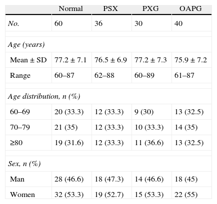
Method

A total of 166 patients were included in this study: 36 eyes with pseudoexfoliation syndrome (PXS), 30 eyes with pseudoexfoliation glaucoma (PXG), 40 eyes with POAG, and 60 normal eyes. Corneal endothelial cell density (ECD), coefficient of variation (CV) in cell size, and percentage of hexagonal cells were measured using a non-contact specular microscope, whereas CCT was measured with an ultrasonic pachymeter.
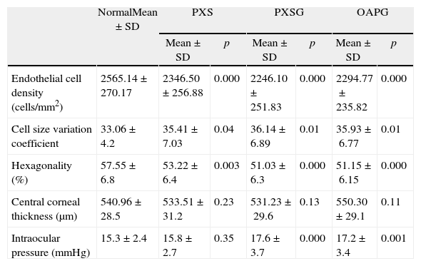
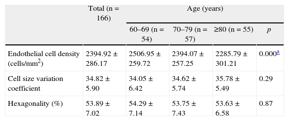
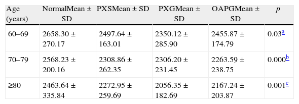
Results

ECD and percentage of hexagonal cells were lower in PEX groups and in the POAG group compared with normal eyes, while the CV in cell size was greater. There was a tendency for greater cell loss and morphological abnormalities of the corneal endothelial cells in PXG eyes compared to PXS eyes, when all pseudoexfoliative eyes were analyzed together. Changes in endothelial cells increased with age. There were no significant differences in mean CCT between the four groups.

Conclusion

Endothelial cell density is significantly decreased, and pleomorphism and polymegathism of cells are increased in PEX eyes, particularly when intraocular pressure is high.

Keywords:
Corneal endothelium
Central corneal thickness
Pseudoexfoliation glaucoma
Specular microscopy
Pseudoexfoliation syndrome
Resumen
Objetivo

Analizar el patrón morfométrico de las células endoteliales de la córnea y el espesor corneal central (ECC) en los ojos con pseudoexfoliativo (PEX) con y sin glaucoma, y compararlos con ojos normales y con glaucoma primario de ángulo abierto (GPAA).

Método

Se incluyeron 166 pacientes en el estudio: 36 sujetos con síndrome seudoexfoliativo, 30 con glaucoma seudoexfoliativo (GPEX), 40 con GPAA y 60 pacientes normales. Los parámetros evaluados con el microscopio especular de no contacto fueron la densidad de células endoteliales (DCE), el coeficiente de variación (CV) del tamaño celular y el porcentaje de hexagonalidad. El ECC se midió con paquimetría de contacto.

Resultados

La DCE y el porcentaje de hexagonalidad celular fueron menores en los sujetos con PEX y GPAA respecto al grupo control, mientras que el CV del tamaño celular fue mayor. Considerando los 2 grupos de ojos con PEX, se observó una tendencia hacia una mayor pérdida de células endoteliales y de modificaciones en los parámetros morfométricos en los ojos con GPEX. Las alteraciones en el patrón especular aumentaron progresivamente con la edad. No hubo diferencias significativas en el valor medio de ECC entre los 4 grupos.

Conclusión

La densidad de células endoteliales está significativamente disminuida y el pleomorfismo y polimegatismo celular incrementado en los ojos con PEX, especialmente cuando la presión intraocular es alta.

Palabras clave:
Endotelio corneal
Espesor corneal central
Glaucoma seudoexfoliativo
Microscopia especular
Síndrome seudoexfoliativo

Article

These are the options to access the full texts of the publication Archivos de la Sociedad Española de Oftalmología (English Edition)
Subscriber
Subscriber

If you already have your login data, please click here .

If you have forgotten your password you can you can recover it by clicking here and selecting the option “I have forgotten my password”
Subscribe
Subscribe to

Archivos de la Sociedad Española de Oftalmología (English Edition)

Purchase
Purchase article

Purchasing article the PDF version will be downloaded

Price 19.34 €

Purchase now
Contact
Phone for subscriptions and reporting of errors
From Monday to Friday from 9 a.m. to 6 p.m. (GMT + 1) except for the months of July and August which will be from 9 a.m. to 3 p.m.
Calls from Spain
932 415 960
Calls from outside Spain
+34 932 415 960
E-mail
Article options
Tools
es en pt

¿Es usted profesional sanitario apto para prescribir o dispensar medicamentos?

Are you a health professional able to prescribe or dispense drugs?

Você é um profissional de saúde habilitado a prescrever ou dispensar medicamentos