


To assess the degree of inter-examiner concordance in the interpretation of periodontal findings in panoramic X-rays in last year students of the School of Dentistry at the University of Colombia.
Material and methodsA descriptive inter-observer concordance-consistency study was conducted for interpretation of periodontal findings in panoramic X-rays among last year students of the School of Dentistry of the University of Colombia and compared results to thos obtained by a periodontics specialist. Concordance evaluation was achieved by means of a kappa coefficient using SPSS Statistics 2.0 program.
Results80 last year students (undergraduate seniors) were evaluated each one assessed two panoramic X-rays, for a total of 160 observations. General concordance established a minimum kappa value of 0.011, and maximum value of 0.720.
ConclusionsConcordance among observing students and the gold standard was poor. This indicated that students did not apply acquired knowledge during their undergraduate dental studies, and provided thus a misleading impression on the circumstances of the periodontium.
Estimar el grado de concordancia inter-examinador en la interpretación de hallazgos periodontales en radiografías panorámicas en estudiantes de último año de la Facultad de Odontología de la Universidad de Cartagena.
Material y métodosSe realizó un estudio descriptivo de concordancia-consistencia inter-observador para la interpretación de hallazgos periodontales en radiografías panorámicas entre estudiantes de último año de pregrado de la Facultad de Odontología de la Universidad de Cartagena, con relación a un especialista en periodoncia. La evaluación de la concordancia se realizó a través del coeficiente kappa utilizando SPSS Statistics 2.0.
ResultadosSe evaluaron a 80 estudiantes del último año de pregrado. Cada uno valoró dos radiografías panorámicas, para un total de 160 observaciones. La concordancia general estableció un valor de kappa mínimo de 0.011 y un valor máximo de 0.720.
ConclusionesLa concordancia entre los estudiantes observadores y el estándar de oro fue pobre. Lo que indica que los estudiantes no aplican los conocimientos obtenidos durante el pregrado de odontología, dando así una errónea impresión del estado periodonto.
Periodontal disease is among the most common infectious diseases found in the human race. It is characterized by inflammation of dental support tissues1–3 including alveolar bone. Diagnosis of this disease is generally emitted based on clinical changes, such as hemorrhage, probing depth, mobility and suppuration without forgetting X-ray findings which in the long term allow us to differentiate among different types of periodontitis.4
Hirschmann5 identified six radiographic findings in periodontium assessment: bone loss, widening of the periodontal ligament space, traumatic occlusion signs, calculi and lamina dura irregularity. The following can also be included: decrease of the alveolar crest height, furcation defects, supra-gingival and sub-gingival plaque retention factors and endodontic-periodontal lesions.6
When assessing periodontal disease7 panoramic X-rays are the most widely tools used as diagnostic help; they possess the advantage of showing all teeth in one single image, inflicting thus low radiation doses to the patient.8,9
Radiographic images provide essential information on bone height, periodontal ligament widening, presence or absence of lamina dura, furcation defects, endodontic-periodontal lesions, plaque retention sites and thus help to determine prognosis and tailor a treatment plan.9–12
Clinical operators constantly reflect variations in the interpretation of diagnostic help and tests.13 Lewis et al14 reported low consensus amongst dentists with respect to study models for occlusal stability and tissue loss in cases of malocclusion. Likewise, Marbach et al15 reported considerable variation amongst clinical instructors in their evaluation of models to assess bruxism severity. Recent research work reported lack of precision and wide variability among dental professionals when evaluating radiographic bone loss. According to the previously exposed, the need arises to propose a research project to assess inter-examiner concordance, so as to generate reflection and discussion on acquired undergraduate dental knowledge.
The present study aimed at showing interexaminer concordance degree in the interpretation of periodontal findings in panoramic X-rays of last year dental students.
MATERIAL AND METHODSThe present article was classified as research lacking biological, sociological of social risk for participant subjects, according to resolution 008430, 4th October 1993, article 11, clause a. Following items described in chapter five of this resolution, all students granted informed consent forms, in said forms they allowed their participation and kept their personal data anonymous. Rejecting participation did not influence the student's academic standing.
Study type: inter-examiner, concordanceconsistency descriptive study undertaken to interpret periodontal findings in panoramic X-rays among last year undergraduate dental students of the Cartagena University comparing them with results obtained by a periodontal specialist.
Population, sample and sample size: 80 last year (senior) undergraduate students participated. Students were enrolled in the 2014 second period of the Cartagena School of Dentistry.
Inclusion criteria: students enrolled in the last (senior) undergraduate year, belonging to the 2007 study plan of the School of Dentistry irrespective or age and gender.
Exclusion criteria: graduated students, students from other faculties and students who declined participation in the present study.
Inclusion criteria for radiographic material: panoramic X-rays (taken in an orthopantomograph brand Veraviewepocs®) of legal-age patients treated at the School of Dentistry of the University of Cartagena.
Data collection and processing: 10 digital panoramic X-rays were randomly selected from the radiographic files of the school of dentistry, for the period June- November 2013. These X-rays were evaluated by a Periodontal Specialist with over 10 years clinical experience. Out of all assessed radiographs, two were selected showing radiographic findings of mild, moderate and severe periodontal disease.
Instrument used for the present study was designed by researchers’ mutual agreement.
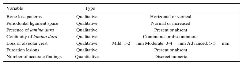
All 80 participants evaluated both panoramic x-rays selected by the gold standard, and they were consigned later in the record format for periodontal findings bearing in mind radiographic parameters (Table I).
Radiographic findings to be evaluated.
| Variable | Type | |
|---|---|---|
| Bone loss patterns | Qualitative | Horizontal or vertical |
| Periodontal ligament space | Qualitative | Normal or increased |
| Presence of lamina dura | Qualitative | Present or absent |
| Continuity of lamina dura | Qualitative | Continuous or discontinuous |
| Loss of alveolar crest | Qualitative | Mild: 1-2mm Moderate: 3-4mm Advanced: > 5mm |
| Furcation lesions | Qualitative | Present or absent |
| Number of accurate findings | Quantitative | Discreet numeric |
Source: Research's own source.
To this effect, all participants possessed the same environmental circumstances, avoiding external distractions, and were allotted a 30 minute time lapse to conduct radiographic assessment; a generic negatoscope, pencil and format to include information were made available to all of them.
Statistical analysis: descriptive analysis of variables was effected with kappa coefficient. This study reflects the degree of concordance among observers, taking values between -1 and +1. The closer the value was to +1, greater would be the degree of concordance among observers. Conversely, the closer the value was to -1 greater would be the inter-observer discordance.16
In order to organize data to be evaluated, a matrix table was achieved, designed in Microsoft Excel, version 2007 for Windows. At a later point, data were assessed with statistical software SPSS Statistics 2.0.
RESULTSConcordance among 80 last year undergraduate dental students was studied, comparing results with those from a periodontics specialist, who was considered the gold standard. Study involved 20 teeth distributed in two panoramic X-rays. Out of total teeth, 10 exhibited some periodontal disease finding and 10 were control cases, for a total of 160 samples.
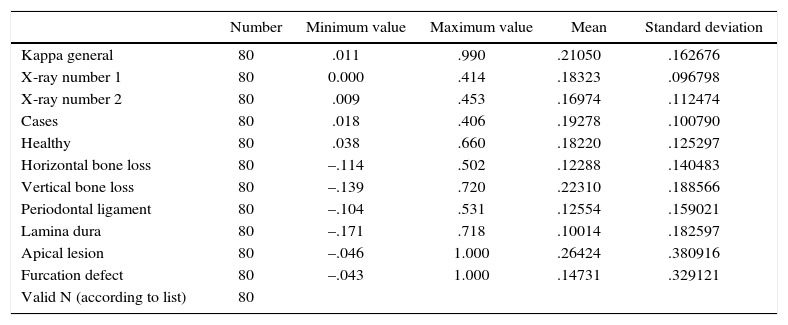
Statistical analysis revealed that the value obtained for kappa general presented a mean of 0.21, with standard deviation of 0.16 (Table II) and was thus interpreted as weak concordance.
Descriptive analysis of all assessed finding.
| Number | Minimum value | Maximum value | Mean | Standard deviation | |
|---|---|---|---|---|---|
| Kappa general | 80 | .011 | .990 | .21050 | .162676 |
| X-ray number 1 | 80 | 0.000 | .414 | .18323 | .096798 |
| X-ray number 2 | 80 | .009 | .453 | .16974 | .112474 |
| Cases | 80 | .018 | .406 | .19278 | .100790 |
| Healthy | 80 | .038 | .660 | .18220 | .125297 |
| Horizontal bone loss | 80 | –.114 | .502 | .12288 | .140483 |
| Vertical bone loss | 80 | –.139 | .720 | .22310 | .188566 |
| Periodontal ligament | 80 | –.104 | .531 | .12554 | .159021 |
| Lamina dura | 80 | –.171 | .718 | .10014 | .182597 |
| Apical lesion | 80 | –.046 | 1.000 | .26424 | .380916 |
| Furcation defect | 80 | –.043 | 1.000 | .14731 | .329121 |
| Valid N (according to list) | 80 |
N: Sample number.
Source: Research's own source.
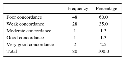
With respect to frequency, table III shows that 60% of all samples reached poor concordance, followed by weak concordance in 35% of all samples.
DISCUSSIONPeriodontal disease is diagnosed with the help of clinical findings. The role of X-rays is aimed to function as a complement, and not a final diagnosis, otherwise, an increase of false positives could occur.9–17
The most common types of X-rays used a diagnosis adjuvant for periodontal disease mainly include panoramic and periapical X-rays.18 The present study was conducted using panoramic X-rays, since they have the advantage over periapical X-rays of showing in one image both jaws with their inserted teeth.2 When a patient is first assessed, it is more common to take panoramic X-rays, nevertheless, when these X-rays show different degree of distortion, it becomes necessary to complement with intraoral X-rays.
Akesson et al10 informed that periapical X-rays were more accurate since they radiographically determined bone height, when compared to panoramic X-ray and bitewing radiographs. Conversely, Gedik et al19 found that periapical X-rays were the less accurate radiographs of the assessed methods. In the present research, participants only interpreted periodontal findings of panoramic X-rays. This fact represents a limitation, since if panoramic X-rays were to be complemented with periapical X-rays, observers could have detected periodontal manifestations with greater accuracy.
When comparing observers and gold standard, exhibited global kappa values were minimum .011 and maximum .990, with mean of 0.21. These data clearly reflect the difference found among participants. Tewary et al,20 found an average of moderate concordance when assessing interexaminer concordance in apical lesions, comparing experienced specialists, specialists in training and oral radiology specialists. They additionally reported that factors which appeared to be of greater impact were the observer's years of experience, it must be equally observed that a minimum one year experience is required in order to obtain moderate concordance. In the present study, although participants were in their last year of dental studies, they did not count with specialized training, and this could explain the low concordance found in the study.
In addition to assessed periodontal findings, it was observed that participants visualized more accurately cases where there is disease than those teeth which are healthy. Apical lesions were the periodontal manifestation with greater degree of concordance.
As already mentioned, a possible explanation could be lack of experience and specialized training, nevertheless, the present study prompts reflection with respect to methodology and learning model provided in the institution with respect to radiographic interpretation and training.
Results of the present study showed little concordance among students and gold standard when comparing periodontal findings in panoramic X-rays. Nevertheless, further studies are required with selected radiographic material and greater image clarity, this would be the case of digital periapical X-rays at different times in order to suitably evaluate progress in the examiners. Therefore it is recommended to initiate students’ valuation, once the use of X-rays as diagnostic adjuvant in clinical practice is established.
Finally, it is suggested to use the methodology developed in this research in other dental areas, to thus achieve uniform criteria among clinical operators.
This article can be read in its full version in the following page: http://www.medigraphic.com/facultadodontologiaunam