


To assess compressive strength of glass ionomer and composite resin restorations in premolar class I cavities.
Material and methodsIn vitro experimental study to assess compressive strength of two types of stomatological restoration materials, using as object of study 52 bi-radicular premolars. Samples were distributed into four groups with different characteristics such as restorative material and cavity depth (2-4mm). Glass ionomer and composite resins were the used restorative materials. Grouped samples were subjected to a compressive vertical force using a EZ-S SHIMADZU texturometer, until achieving the material's fracture. Obtained data were subjected to the Shapiro-Wilk test in order to assess data normalcy, null hypothesis was rejected. Total data analysis was conducted with t-Student test for independent samples.
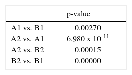
ResultsData obtained after assessing superficial hardness of different restorative materials showed the existence of statistical differences which favored composite resin when compared to glass ionomer at both depths (p = 6.908 × 10⁻¹¹ and p = 0.000). In intra-group comparison, a significant different was found between both groups (resin and glass ionomer) at different depths (p = 0.000155887 and p = 0.00257443).
ConclusionAssessment of 4mm tooth cavities restored with Tetric N-Ceram resin revealed greater hardness than those accomplished with Vitremer® resin at 2 and 4mm and with the same resin at 2mm depth.
Evaluar la resistencia a la compresión en restauraciones de ionómero de vidrio y de resina compuesta en cavidades clase I en premolares.
Material y métodosUn estudio experimental in vitro, para evaluar la resistencia a la compresión de dos tipos de materiales restaurador estomatológico, utilizando como objeto de estudio 52 dientes premolares birradiculares. Las muestras fueron distribuidas en cuatro grupos con diferencias en sus características, como fueron el material restaurador y la profundidad de la cavidad (2-4mm). Se empleó como material restaurador ionómero de vidrio y resina compuesta. Las muestras grupales fueron sometidas a una fuerza vertical compresiva utilizando un texturómetro EZ-S SHIMADZU hasta lograr producir la fractura del material. Para evaluar la normalidad los datos obtenidos se sometieron a la prueba Shapiro-Wilk que rechazó la hipótesis nula. El análisis de los datos totales se realizó a través del test t-Student para muestras independientes.
ResultadosLos resultados obtenidos al evaluar la dureza superficial de los diferentes materiales restauradores, muestran que existen diferencias estadísticas a favor de la resina compuesta en comparación con el ionómero de vidrio en ambas profundidades (p = 6.908 × 10⁻¹¹ y p = 0.000), y en la comparación intragrupal se aprecia una diferencia significativa entre los dos grupos de resina e ionómeros a distinta profundidad (p = 0.000155887 y p = 0.00257443).
ConclusiónAl evaluar las cavidades de los órganos dentarios de 4mm de profundidad, que fueron restaurados con resina Tetric N-Ceram, éstas presentan mayor dureza en comparación con los que fueron restaurados con resina VitremerTM a 2 y 4mm y que la misma resina a 2mm de profundidad.
Tooth degeneration is caused by different factors which can affect tooth's enamel, dentin and hard tissues.1 If said degenerative process is found at an initial stage, it can be reversible, such is the case of the whitish spot; if this is not the case, an irreversible process sets in related to the cavitation presence. For these reasons, to count with ideal and longer-lasting materials is of the utmost importance when restoring original cavities caused by different carious processes in the mouth.2
Several research projects are proof that the scientific community is interested in improving mechanical properties of filling materials, remembering nevertheless that there are still some deficiencies such as low resistance to wear, micro-filtration, pigmentation and incomplete polymerization. Resistance of these materials to diverse factors is still not ideal and results in their short permanence in the mouth, nevertheless, some of these materials have proven to possess annual wear similar to that of silver amalgam.3,4
It is important to bear in mind some current and relevant concepts of minimally invasive dentistry: when teeth require restoration, this restoration must be as conservative as possible with the dental structure when required preparations are undertaken. This has caused abandonment of certain materials requiring extensive preparations in order to acquire resistance and adhesion to the tooth. Contrarily, the use of materials not requiring extensive preparations to be used in different cases is on the rise.5
Certain characteristics of the material provide confidence to the clinical operator, who will play an important role when choosing materials. These characteristics, among others, are resistance to masticatory forces, acceptable esthetics, and superficial hardness.6
In a publication previous to this study, Taron et al, in 2015, proposed in a pilot study as experimentation model a large number of natural teeth previously extracted due to orthodontic reasons. This sample was used to develop fracture resistance and tolerance tests. The study nevertheless demanded evidence of sample increase and model refinement.6
Restorative materials presently used such as composite resin and glass ionomers, possess advantages and disadvantages, therefore the aim of the present study was to compare one of the multiple characteristics essential to restorative materials, that is to say compressive strength of the aforementioned two materials.
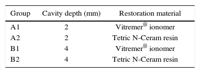
MATERIAL AND METHODSAn in vitro quasi-experimental study was conducted. In it, assessment was made of compression resistance of a reconstructive glass ionomer in contrast to a nano-hybrid composite resin, both materials were used to restore Black's class I cavities with depths of 2 and 4mm in human premolars. The convenience-selected sample was composed of 52 premolars, extracted during orthodontic treatments, lacking extensive enamel anomalies.
The sample was divided into two groups: group A, for teeth where 2mm deep cavities were performed, and group B where 4mm deep cavities were established. A blunt edge, cylindrical diamond burr was used. Depth of all prepared cavities was rectified with a millimeter periodontal probe (Hu-Friedy).
A self-polymerizing acrylic support was manufactured for each tooth in the sample, so as to provide stability when positioned in the compressive strength measuring instrument.
Both groups were divided into two sub-groups. Number 1 was for teeth used as sample, restored with reconstructive glass ionomer, brand 3M Vitremer®. Number 2 was for teeth restored with nano-hybrid resin Tetric N-Ceram, brand Ivoclar Vivadent (Table I).
All teeth of the sample were subjected to stress tests with texturometer EZ-S SHIMADZU, series number 346-54909-33, with 50-60Hz, with maximum capacity range of 500 Newton. Filled and restored teeth were subjected to compression in the occlusal side, with a 1mm contact area, until achieving a 1mm depth in one single advance (Figure 1). Strength necessary to monitor necessary strength to penetrate in the vertical aspect of the restoration existing in all teeth was monitored. It must be stressed that in all samples force application was equally performed at the central point of the restoration.

Ethical considerations of this project were in concordance with resolution 008430 (1983), Ministry of Health, Colombian Republic.
Statistical analysisA matrix table was manufactured from obtained results, to this effect Microsoft Excel version for Windows 7 was used. After this, the Shapiro-Wilk test was applied to each of the samples. The following results were obtained: A1 = 0.059, A2 = 0.940, B1 = 0.987 and B2 = 0.300. Since values were above 0.05, normalcy hypothesis could not be discarded. This test was conducted with program SPSS Statistic v22 IBM. T-Student test was applied for independent samples, with significance level p > 0.05, using Statgraphics portable program centurion XV.II.
RESULTSAfter Applying t test for independent samples analyzed two by two, it was found that cavities measuring 2mm and filled with Vitremer® and those filled with Tetric N-Ceram exhibited significant differences (p = 0.00000000006908). A 95% confidence interval was obtained for mean differences, supposing equal variances (-60.0973 up to -41.1631). Since confidence interval does not contain 0 there was a statistically significant difference between means of both samples, with a 95% confidence level. Tested resin exhibited greater significance, since it possessed greater mean (419.9500) as observed in figure 2.

Figure 3 shows results of the comparison of both used restorative materials, after conducting an analysis of the 4mm cavities filled with Vitremer® and Tetric N-Ceram resin. They exhibited significant difference (p = 0.000) and a confidence interval comprised between values of -94.8257 up to -76.604. Due to the fact that confidence interval does not contain 0, there was a statistically significant difference with confidence interval of 95%. Resin was more significant since it possessed greater mean (438.9784N).

When comparing 2 and 4mm cavities filled with Vitremer®, results showed no statistically significant difference (p = 0.00257443), confidence interval 6.07823 up to 25.3742, since interval contains 0 no statistically significant difference was found between both samples, with a 95% confidence level. Comparison with 2 and 4mm cavities filled with Tetric N-Ceram resin, results revealed statistically significant difference (p = 0.000155887), alternatively, appealing to the confidence interval (-28.2774 up to -10.4401); since interval did not contain 0 there was a statistically significant difference between means of both samples, with confidence interval of 95%. Significance of resin at 4mm was greater since it possessed greater media (438.9784N) (Tables II and III).
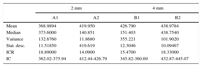
Descriptive statistics. Comparison of superficial hardness of restoration materials at different depths.
| 2 mm | 4 mm | |||
|---|---|---|---|---|
| A1 | A2 | B1 | B2 | |
| Mean | 368.9894 | 419.950 | 426.790 | 438.9784 |
| Median | 373.6000 | 140.851 | 151.403 | 438.7540 |
| Variance | 132.6760 | 11.8680 | 355.221 | 101.9020 |
| Stat. desc. | 11.51850 | 419.619 | 12.3046 | 10.09467 |
| ICR | 18.89000 | 14.0900 | 15.4700 | 18.33000 |
| IC | 362.02-375.94 | 412.44-426.79 | 345.82-360.69 | 432.87-445.07 |
In a previous publication of this research group, the cavity model was primed in natural teeth, in order to try to establish the importance of research in an environment much more similar to the reality of the oral cavity in human beings.6 Irrespectively of cavity depth, greater superficial hardness could be observed in all teeth restored with nano-hybrid resin. Nevertheless, in all study groups resin placed in 4mm deep cavities exhibited higher hardness. Group B1 exhibited lowest results upon penetration.
Carrillo (2008)7 reported similar results to those obtained in the present study with respect to resistance of some filling materials used in dentistry. This study reported comparison of composite resin, reconstructive glass ionomer and fluid resin; in it, hardness values of composite resin were widely greater than those of the two remaining materials.8,9
In 2014, Suarez and Lozano10 studied hardness of different types of resins, but, differing from the present study, they conducted their study examining the material in the shape of pre-formed elements, built with the studied materials, and not in a tooth by filling directly a prepared cavity, mimicking thus clinical reality. It is considered that the model proposed in the present study, far more resembles a real scenario of resistance measurement and compressive forces.
Sun Ae Song et al, in 201411 conducted research on resin hardness at different polymerization stages. Nevertheless, they conducted that research with Vickers’ microdurometer, which differs from the texturometer using in the present study, since its measurement is not directed to assess what force is needed by the machine in order to achieve penetration.12
To conduct studies on assessment of superficial hardness of two dental filling materials at two different thicknesses or depths is very important for the industry of dental materials, and for modern dentistry since contributions achieved with these research projects help to refine clinical indications and guide dental materials manufacturers in the search for further benefits for dental patients. This point was taken by Shanthala (2013)12 and Erazo (2010)13 since they considered this a series of factors which allowed to achieve longer and more effective dental treatments for patients in cases when glass ionomer or resins were used as filling materials.14,15
CONCLUSIONBearing in mind limitations inherent to an in vitro study, it could be concluded that teeth with 4mm deep cavities restored with Tetric N-Ceram exhibited greater hardness than those restored with Vitremer® at 2 and 4mm and 2mm deep cavities with the same resin, nevertheless it must be accepted that evolution of present research might alter these results.
Resistance to compressive strength showed that to restore posterior teeth, studied resin possessed significantly higher hardness when compared to reconstructive glass ionomer.
Range of obtained forces by no means compare to range of forces recorded in human teeth bite. This points out to the need to improve presently used dental materials.
Bearing in mind diverse applications of used methods and materials, it would be possible to create new research projects targeting changes in hardness of restorative materials.
This article can be read in its full version in the following page: http://www.medigraphic.com/facultadodontologiaunam
DDS, School of Dentistry, University of Cartagena.
Pharmaceutical Chemist, University of Cartagena. Degree in Biotechnology, University of La Habana. PhD Candidate in Food Science, University of La Habana. Professor, University of Cartagena.
DDS, University of Cartagena. Orthodontics Specialist, University of Saõ Paulo. Degree in Applied Statistics, Universidad del Norte. Professor, University of Cartagena.
DDS, University of Cartagena. Periodontics Specialist, Universidad Javeriana. Degree in Education, Universidad del Norte. PhD in Biomedical Sciences, University of Cartagena. Professor, University of Cartagena.