


Recent publications done in well-recognized journals of orthodontics, assure that micro-osteoperforations (MOP) enhance the process of bone remodeling and accelerate dental movement. Its application is easy, fast and can be performed by the orthodontist.
ObjectiveThe objectives are to evaluate and compare canine distalization time in young patients, by using both an acceleration technique through MOP and a conventional technique in a split-mouth design. 1. Find the best procedure to accelerate an orthodontic treatment with extractions. 2. Decrease treatment time.
Material and methodsCanine distalization was performed on 10 young patients whose treatment plan included first premolar extractions. MOP was performed in the first premolar extraction zone of the right quadrant. A mini-implant was placed between the second premolar and the first molar in order to obtain absolute anchorage. Traction was applied by using power chains on both sides. In the left quadrant canine distalization was performed through conventional methods.
ResultsThe obtained results in the side where MOP was performed, show a significant reduction in the procedure time when compared with conventional treatment.
ConclusionsCanine distalization acceleration using MOP in patients with extractions leads to highly effective results; up to 41% faster space closure. This results in a shorter and more comfortable orthodontic treatment for the patient.
Diferentes investigaciones aseguran que las micro-oseoperforaciones (MOP) aumentan el proceso de remodelado óseo y aceleran el movimiento dental, su aplicación es fácil, rápida y la puede realizar el ortodoncista.
ObjetivoLos objetivos son evaluar y comparar el tiempo de distalización canina en pacientes jóvenes con la técnica de aceleración mediante MOP y técnica convencional en boca dividida; y encontrar el mejor procedimiento para acelerar un tratamiento de ortodoncia con extracciones y disminuir el tiempo de tratamiento.
Material y métodosSe llevó a cabo la distalización de caninos en 10 pacientes jóvenes cuyo plan de tratamiento incluía extracción de primeros premolares, se practicó MOP en la zona de la extracción del primer premolar del cuadrante derecho, se colocó un mini-implante entre el segundo premolar y primer molar para obtener un anclaje absoluto, haciendo la tracción con cadena elástica en ambos lados, en el cuadrante izquierdo la distalización de canino se llevó a cabo con procedimiento convencional.
ResultadosEl resultado que se obtuvo en el lado que se practicó MOP muestra una disminución considerable en el tiempo empleado para este procedimiento comparado con el método convencional.
ConclusionesAcelerar la distalización canina con micro-oseoperforaciones en pacientes con extracciones resulta muy eficaz hasta un 41% de cierre de espacios más rápido y con ello un tratamiento de ortodoncia más corto y cómodo para el paciente.
The main concern of patients before starting orthodontic treatment is how long it will take to complete the orthodontic treatment.
The main setback occurs in treatment of class II patients who require first premolar extractions to distalize the canines. This procedure leads to a longer treatment time.
In the last few years several devices and treatment options have made the orthodontic process more efficient and comfortable but not faster.
At present new techniques have been introduced to accelerate tooth movement such as corticotomies and application of prostaglandin E2, but these procedures are painful and costly since they require another specialist to perform them. However, techniques of micro-osteoperforations have been developed recently which increase the process of bone remodeling, have a quick and easy application and can be performed by the orthodontist without pre-surgical discomfort.
BACKGROUNDThe main resistance to tooth movement was the cortical plates of the bones and the interruption of its continuity. The procedure involves the reflection of total thickness flaps to expose buccal and lingual alveolar bone, followed by the interdental cuts through cortical bone and barely penetrate into the medullary bone to accelerate orthodontic movements.1–3
Duker used Kole's basic technique in dogs to investigate if the cuts affected dental vitality. It was concluded that neither the pulp nor the periodontal tissues were damaged during orthodontic tooth movement after the corticotomy and interdental cuts were performed.4
Frost in 1983 described the regional acceleratory phenomen (RAP) as the local response to a noxious stimulus. He described a process by which a tissue is formed faster than the normal regional regeneration process. Through the improvement of the various stages of healing, this phenomenon makes scarring occur 2-10 times faster than normal physiological healing.5,6
Arias and Márquez Orozco in 2006, based on these findings formulated the hypothesis that the limited and shallow drilling of the buccal or labial cortical plate of the maxilla would be sufficient to increase the expression of inflammatory cytokines, speeding up the process of bone remodeling and therefore the speed of tooth movement.2
Mani Alikhani et al. in 2013 stated that micro-osteoperforations are an effective, safe and comfortable procedure that accelerates tooth movement significantly and could result in shorter orthodontic treatments.7 They mention that this new minimally invasive technique stimulates the activity of cytokines to accelerate the remodeling of the alveolar bone.
The regional acceleration phenomenon f (RAP) begins from the lesion with a maximum acceleration and declines using corticotomies. RAP begins within days of the injury and it usually reaches its peak at 1-2 months. It usually lasts 4 months in bones and may take from 6 to 24 months and more to disappear.8 Acceleration has been used primarily in patients with biprotrusion and skeletal anchorage to distalize the anterior segments.9
Different techniques have been used to achieve faster tooth movement. Throughout history, there have been studies of different methods: prostaglandins, corticosteroids, hormones, electric currents and magnetic fields, mechanical vibration, corticotomies and micro-osteoperforations. 7,10–19 By combining corticotomies and skeletal anchorage, better results are achieved without secondary movements.20,21 This serves as well for the distalization of segments.22
Linkow in 1969 described for the first time the use of implants as orthodontic anchorage in patients, by using them for retraction of the upper anterior teeth. Several authors have used them as well to perform different movements.23–27 Kuroda in 2009 determined that for canine distalization using TADs between the roots of the second premolar and first molar a NiTi spring with 100 grams of force must be used.28 In the mandibular arch min-screws may be used in patients with biprotrusion.29
Mini implants placed at 90o may be a better option in orthodontic treatments due to a greater resistance to forces and therefore, more stability.30
Giuliano Maino in 2013 described an anchoring system with self-drilling titanium mini-screws with immediate loading called Spider Screw TM. This anchoring system may be used for different types of orthodontic movements due to the immediate loading using between 50 and 250 g. Mini-screws do not have osseo-integration, which facilitates their removal.31,32
CASE REPORTDiagnosisA female patient of 14 years of age was referred to the Orthodontics Clinic of the Faculty of Dentistry, Torreon Unit of the Universidad Autónoma de Coahuila for presenting moderate crowding and lack of eruption of an upper canine. While performing the medical and dental history it was found that the health status of the patient was apparently healthy, no pathological data were observed during the oral examination; she did not have pain, or any manifestation of temporomandibular joint disorders.
The analysis of the facial photographs revealed a dolichofacial patient, oval face, straight nose, biprocheilia and coincident facial and dental midlines (Figures 1A-1C).

Upon the intraoral clinical examination it was observed: lower midline deviated to the left, healthy gum, right upper canine in supra-eruption, 3 mm overbite, 3 mm overjet, molar class II, canine class II and oval arch form (Figures 2A-2E).

In the orthopantomography, symmetrical mandibular condyles and ramus were observed, continuous alveolar ridges, 28 erupted teeth, the germ of the third molars and second premolars as well as a crown-root ratio of 2:1 (Figure 3).

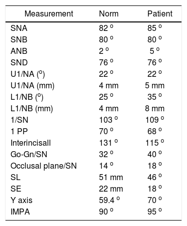
The cephalometric data revealed a class II due to maxillary hyperplasia, vertical growth, tendency to skeletal open bite, short anterior cranial base, short mandibular ramus and posterior position of the mandible; dental class II, lower incisor proclination, lower incisor protrusion, tendency to open bite, increased lower third and oval face (Figure 4andTable I).

Cephalometric data.
| Measurement | Norm | Patient |
|---|---|---|
| SNA | 82 o | 85 o |
| SNB | 80 o | 80 o |
| ANB | 2 o | 5 o |
| SND | 76 o | 76 o |
| U1/NA (o) | 22 o | 22 o |
| U1/NA (mm) | 4 mm | 5 mm |
| L1/NB (o) | 25 o | 35 o |
| L1/NB (mm) | 4 mm | 8 mm |
| 1/SN | 103 o | 109 o |
| 1 PP | 70 o | 68 o |
| Interincisall | 131 o | 115 o |
| Go-Gn/SN | 32 o | 40 o |
| Occlusal plane/SN | 14 o | 18 o |
| SL | 51 mm | 46 o |
| SE | 22 mm | 18 o |
| Y axis | 59.4 o | 70 o |
| IMPA | 90 o | 95 o |
To achieve canine class I, molar class I, to improve the overjet and overbite, to obtain matching dental midlines, to improve the profile.
Treatment planThe treatment plan included the extraction of four first premolars, leveling, distalization of the upper canines, retraction of the upper and lower anterior segments and finishing of the case.
For distalization of the upper canines it was suggested to perform micro-osteoperforations to shorten the time of this procedure and to compare the effectiveness of the micro-osteoperforations to accelerate orthodontic movement. The study was done with a split-mouth design. The patient accepted this proposal signing the informed consent. The treatment was performed in the Clinic of the Masters in Dental Sciences with Emphasis in Orthodontics in the Faculty of Dentistry, Torreon Unit of the Universidad Autónoma de Coahuila.
Treatment progressThe patient had the extraction of four first premolars. During this event, the patient's parents read the informed consent and signed the authorization. 0.018” slot Alexander brackets were placed, alignment and leveling was achieved, as well as to position the upper right canine within the arch to distalize it. 0.018” slot Alexander bands were placed and also, a TPA for anchorage.
To increase the certainty of the distal displacement of the canines mini-implants were used as anchorage. The inter-radicular distance between teeth #1.5 and 1.6 was assessed radiographically to determine the dimensions of the mini-implant that was going to sustain the strain.
The mini-implant was placed and one week after micro-osteoperforations were conducted.
Topical anesthetic was applied at the extraction site and left there to act for 5 minutes. This procedure allows the patient to report any discomfort as well as to detect contact with some root during mini-implant placement. Three micro-osteperforations were performed in the cortical bone at the extraction site.
Elastomeric chains were placed from the mini-implant to the bracket of tooth #1.3 with a force of 150 g (Figure 5). Another elastomeric chain was placed from teeth #2.3 to 2.6 with a force of 150 g using a 0,016” stainless steel archwire.

Before activation, the distance between distal of the canine and mesial of the premolar was measured with a digital caliper (Figures 6A-6C). Appointments were indicated every 15 days in order to maintain the force of the chain (Figures 7 to 13). Distalization time with micro-osteoperforations was 10 to 12 weeks.
RESULTS


The results were obtained with the analysis and comparison of the photographs and measurements with the digital caliper appointment after appointment (Figures 14and15).


The result was evaluated at the end of the distalization period of both quadrants. Complete distalization was achieved more effectively and rapidly in the quadrant of MOP in comparison with the control quadrant (Figure 16).

Measurements of the remaining space in the area of distalization measured every two weeks with digital caliper. The measurements were compared between the quadrants of micro-osetoperforations and controls, obtaining an average of 45.20% of greater effectiveness in distalization with MOP than the control.
The efficiency, simplicity and low cost of micro-osteoperforations have been some of the advantages provided by this technique to simplify accelerated orthodontics, being increasingly innovative and comfortable as well as within the reach of all orthodontists thanks to the ease for performing them and their low cost.
The literature reports several methods of accelerated orthodontics. Most of them describe multiple cases in both animals and humans with excellent results; the majority of these studies submit the patient to more complex surgical procedures in which another specialist must intervene, either the maxillofacial surgeon or the periodontist, in order to perform them, thus increasing costs and posttreatment care.
Kole in 1949 1 through their research on interproximal cuts in the bone cortex reported that they were highly effective in accelerating tooth movement and in obtaining satisfactory results. The disadvantage was that, at the time, the technique was very aggressive, was not well accepted and its posttreatment complications were a problem to consider.
Duker in 19753,4 described in their results that neither the pulp nor the periodontal tissues were damaged during corticotomies using the technique established by Kole,1 this is one of the main advantages of the cuts or perforations of bone. To achieve these results, in the technique hereby described, we decided not to apply local anesthesia, but only topical anesthesia to preserve the sensitivity of the periodontium and teeth, hence, if the perforation performed by the orthodontist was very close to the periodontium, the patient would inform at the time of discomfort giving the specialist a chance to redirect the perforation.
Mani Alkahani et al. in 20137 achieved highly favorable results for tooth movement doing micro-osteoperforations in rats and afterwards, in humans in which the result was not so surprising as in rats but similarly accelerated tooth movement after the implementation of MOP. By making a split-mouth study in the case of one patient, a more rapid movement was observed in comparison to a normal technique. These results have great impact, since they come from the same patient, thus we may assess optimal and reliable results by performing both techniques in the same host.
Taking into consideration the recommendations described the least invasive method was chosen in order to motivate a patient who was reluctant to the extractions needed for orthodontic treatment. It was decided to make a split-mouth comparison in the absence of studies conducted in a same patient to achieve highly reliable results retracting the canine through a technique with 0.022 slot Alexander brackets using the same force on both sides, On the control the side, anchorage was increased with a mini-implant between teeth #1.5 and 1.6 to achieve only canine distalization, avoiding any mesialization of the posterior segment due to bone softening produced with MOP. Both sides received a load of 150 g.
Several authors promote the use of mini-implants for anchorage in upper anterior movements.23–27 The force established by Kuroda in 200928 for canine distalization with a mini-implant between teeth #1.5 and 1.6 was 100 g with a closed NiTi coil spring. The grams used in this technique for both sides were 150 g. Due to the fact that the traction was performed with elastomeric chains and that they tend to lose their strength as time goes by, it was preferred to apply 150 g of force and renew the chains every 15 days to preserve the desired force.
Giuliano Maino and other authors in 201329–31 preset that the load on mini-implants can be immediate if and when the forces are between 100 to 200 grams. Using the results indicated by these authors the decision to perform immediate loading were taken. The results were favorable without complications thanks to the prior study of cases of anchorage with these factors.
CONCLUSIONSpace closure through canine distalization with the aid of micro-osteoperforations is a less invasive option for accelerated orthodontics. It decreases the risks in comparison with other techniques such as corticotomies, in addition to being a treatment available to more patients with economic constraints.
The success or failure of the technique will depend on the specialist, mainly at the time of MOP. It is necessary to consider that bone density varies between each patient, as well as the care and perseverance that the patient has during treatment.