


Hand-wrist radiographs have been used to determine the variations of skeletal maturation by many researchers. The morphology of cervical vertebrae has been used to assess skeletal maturation and its possible relationship with facial growth. The aim of the following article was to determine the correlation between the growth stages in the cervical and hand-wrist bone maturation analysis in Mexican boys and girls ages 9 to 16 years at the Children's Hospital of Mexico «Federico Gomez» during 2011.
MethodologyLateral headfilms and hand-wrist radiographs from 194 files of the Orthodontics Department of the Children's Hospital of Mexico «Federico Gomez» were examined. The exclusion criteria were: patients with craniofacial syndromes, growth disorders and metabolic disorders that affected growth. The values obtained for skeletal maturation from both analysis were analyzed through the SPSS v.17.0 (Statistical Package for the Social Sciences) software using the Spearman test to study the correlation between the two analyses.
ResultsA high correlation (0.89) was found between the Fishman's hand-wrist analysis and the Lamparski cervical analysis. It was also found that the largest correlation by age was at the age of 9 (0.94). The smallest correlation by age was at 16 years old (0.68). The average age was 12.7 years.
ConclusionsLateral headfilms are a valuable auxiliary to predict the growth spurt. In some cases, it may not be necessary to obtain a hand-wrist radiograph since the information of the vertebrae might be sufficient to determine the skeletal maturation stage.
Las radiografías de mano-muñeca han sido usadas para conocer las variaciones de maduración ósea, de muchas formas y por muchos investigadores; así como también para valorar la maduración esquelética y la posible relación con el crecimiento facial se ha usado la morfología de las vertebras cervicales. El objetivo del siguiente artículo fue determinar la correlación existente entre los estadios de crecimiento en los análisis cervical y carpal de maduración ósea en niños y niñas mexicanos de 9 a 16 años del Hospital Infantil de México «Federico Gómez» en el 2011.
MetodologíaSe examinan radiografías lateral de cráneo y carpales de 194 expedientes del Servicio de Ortodoncia del Hospital Infantil de México «Federico Gómez» entre las edades de 9 a 16 años. En los criterios de exclusión fueron: pacientes sindrómicos con involucración craneofacial, trastornos en el crecimiento, trastornos metabólicos que afecten el crecimiento. Se analizan los valores obtenidos de maduración ósea de ambos análisis a través del programa SPSS v.17.0 (Statistical Package for the Social Sciences), utilizando la prueba de Spearman para estudiar la correlación de los dos análisis.
ResultadosSe encontró una alta correlación de 0.89 entre el análisis carpal de Fishman y el análisis cervical de Lamparski. De este modo también se halló que la mayor correlación por edad fue a los 9 años con 0.94. Así como la correlación menor por edad fue a los 16 años con 0.68. El promedio de edad que se obtuvo fue de 12.7 años.
ConclusionesLa radiografía lateral de cráneo es un valioso auxiliar para predecir el pico de crecimiento. En algunos casos puede no ser necesario mandar a tomar una radiografía carpal y puede ser suficiente la información de las vértebras para establecer el estadio de maduración ósea.
Each person matures in his or her individual time and this is the case where the value of the hand-wrist radiograph becomes evident. Hand-wrist radiographs have been used for this purpose in many ways and by many researchers.
Individuals who have shown delay or acceleration in their maturation stages exhibit delays or accelerations comparable to facial and bone growth.1
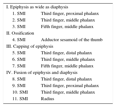
Fishman hand-wrist analysis. Skeletal maturation assessment systemThe sequence of the four stages of ossification (Table I) progresses through the breadth of the epiphyses in specific phalanges, the ossification of the adductor sesamoids of the thumb, the lining of the specific epiphyses over the epiphyses, the fusion of specific epiphysis and the epiphysis equal in width to the diaphysis.1
Four ossification stages and the eleven skeletal maturation indicators that compose the Fishman's hand-wrist analysis.
| I. Epiphysis as wide as diaphysis | |
| 1. SMI | Third finger, proximal phalanx |
| 2. SMI | Third finger, middle phalanx |
| 3. SMI | Fifth finger, middle phalanx |
| II. Ossification | |
| 4. SMI | Adductor sesamoid of the thumb |
| III. Capping of epiphysis | |
| 5. SMI | Third finger, distal phalanx |
| 6. SMI | Third finger, middle phalanx |
| 7. SMI | Fifth finger, middle phalanx |
| IV. Fusion of epiphysis and diaphysis | |
| 8. SMI | Third finger, distal phalanx |
| 9. SMI | Third finger, proximal phalanx |
| 10. SMI | Third finger, middle phalanx |
| 11. SMI | Radius |
Lamparski created a series of standard values for vertebral maturation. He reported that his series of standard values was as accurate as the hand-wrist method for assessing skeletal maturation. Subsequently different authors have reported the relationship between CVM (Cervical Vertebrae Maturation) and skeletal maturation as compared to hand-wrist radiographs. It has also been reported a correlation between CVM mandibular and growth.
The CVM method has been used as a tool to help determine the ideal time for orthopedic treatment. Bacceti provided clinical protocols for treatment of the malocclusions in three dimensions. These protocols require a strict and accurate identification of the stages of the CVM to be clinically applicable. If they are accurate and clinicallly reproducible, these protocols will give the clinicians a valuable and reliable assessment of their patients’ growth with a routine radiograph: the lateral cephalogram. However, before the clinical use of the CVM method is accepted, its accuracy and reproducibility should be assessed using methods that eliminate the inconveniences in the methodology of the previous studies.2
MeasurementAfter completion of endochondral ossification, vertebral growth takes place by apposition in the periosteum. It seems to take place only frontally and laterally. Todd et al and Lanier et al performed measurements in lateral headfilms of the lower cervical vertebrae. Lamparski studied changes in size and shape of the cervical vertebrae to create standard values of maturation of the cervical vertebrae. His method was taken from Todd and Pyle, Elsber and Dyke, Lanier, Bick and Copel and Hinck.3
METHODOLOGYThe present study was carried out during 3 months at the Children's Hospital of Mexico «Federico Gomez», in the Department of Stomatology and Specifically in the Orthodontics Department. The observation units were the hand-wrist radiographs and lateral headfilms. A non-probability sampling method was used and 194 rays were observed.
The universe of study included patients who met with the following inclusion criteria: patients of Mexican descent, with complete records, who had been referred to the Orthodontics Department for treatment, were in an age range of 9 to 16 years old and had their hand-wrist radiographs and lateral headfilms taken. The exclusion criteria were: patients with congenital syndromes that presented craniofacial involvement, patients with growth disorders (for example dwarfism or gigantism) or with metabolic disorders that affect the growth (for example hypothyroidism or diabetes mellitus).
RESULTSThe study consisted in the analysis of 194 lateral hand-wrist radiographs and lateral headfilms using the Fishman analysis for hand-wrist radiographs, which includes 11 indicators also called SMI (Skeletal Maturational Indicators). For the lateral headfilms the Lamparski analysis was used which includes 6 stages also called CVMI (Cervical Vertebrae Maturation Index).
The sample mean was 12.7 years with a minimum age of 9 years and a maximum age of 16 years (Figure 1). The interval for the 9-16 years sample was 7.

Patient distribution according to age. In the age distribution according to frequency it was observed that the ages with a larger frequency are found in the centre of the graph, at the age of 12 and 13 years and the lower-frequency are at the ends, as well as the age range that is 9 to 16 years.
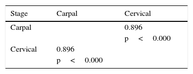
In the Spearman test there was a correlation of 0.89 between the carpal and cervical stages which was statistically significant (Table II).
Rho Spearman correlation analysis between the skeletal maturation stages.
| Stage | Carpal | Cervical |
|---|---|---|
| Carpal | 0.896 | |
| p<0.000 | ||
| Cervical | 0.896 | |
| p<0.000 |
The correlation was significant at a 0.01 level (bilateral).
The obtained correlation was 0.89 between the carpal and cervical stages which indicates a high correlation between them.
In the analysis of the hand-wrist radiographs the indicator with the highest frequency was 49 in the MP5 Cap (SMI 7) stage and the lower frequency was 1 which is the PP3 widening (SMI 1). It was also found that the largest number of cases in the carpal indicator was in MP5 Cap (SMI 7) with 46 cases and the least amount of cases in the carpal indicator was in DP3 Cap (SMI 5) Ru (SMI 11) with 4.
On the other hand, in the analysis of the cervical radiograph sample, it was observed that the higher stage frequency was 72 upon deceleration (CVMI 4) and the lower frequency was 4 upon completion (CVMI 6). The least amount of cases in the cervical analysis was upon Initiation (CVMI 1) with 13 cases.
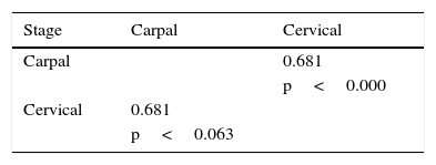
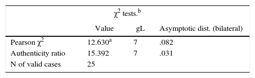
Based on the above-mentioned test it was found that the highest correlation by age was at 9 years old with 0.94 (Table III). The lowest correlation by age was 16 years with 0.68 (Table IV). There was a marginal difference at the age of 10 with 0.92 and the minimum expected frequency was 0.25 (Table V), as well as a marginal difference of 0.82 at the age of 11. The minimum expected frequency was 0.36 (Table VI).
High correlation by age between cervical and carpal analysis.
| Stage | Carpal | Cervical |
|---|---|---|
| Carpal | 0.949 | |
| p<0.000 | ||
| Cervical | 0.949 | |
| p<0.000 |
The highest correlation by age was at 9 years with 0.9 which is a very close figure to 1.00 that represents a total correlation between the cervical and carpal analysis.
Low correlation by age between cervical and carpal analysis.
| Stage | Carpal | Cervical |
|---|---|---|
| Carpal | 0.681 | |
| p<0.000 | ||
| Cervical | 0.681 | |
| p<0.063 |
The lowest correlation by age was at 16 years with 0.68 representing the stage group in which the least amount of coincidences were observed between the carpal and cervical analysis.
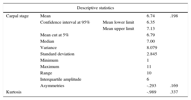
During the analysis of the hand-wrist radiograph of the Fishman analysis 11 indicators, a mean of 6.74 was obtained with indicator number 6 as the mean value (Table VII).
Fishman analysis.
| Descriptive statistics | ||||
|---|---|---|---|---|
| Carpal stage | Mean | 6.74 | .198 | |
| Confidence interval at 95% | Mean lower limit | 6.35 | ||
| Mean upper limit | 7.13 | |||
| Mean cut at 5% | 6.79 | |||
| Median | 7.00 | |||
| Variance | 8.079 | |||
| Standard deviation | 2.845 | |||
| Minimum | 1 | |||
| Maximum | 11 | |||
| Range | 10 | |||
| Interquartile amplitude | 6 | |||
| Asymmetries | -.293 | .169 | ||
| Kurtosis | -.989 | .337 | ||
Fishman analysis, a mean of 6.74 was obtained with indicator number 6 as the mean value.
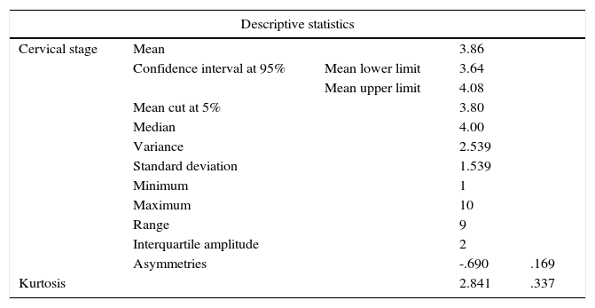
In the statistical analysis of the Lamparski cervical radiograph analysis a 3.86 mean was obtained with stage 3 as the mean value (Table VIII).
Lamparski analysis.
| Descriptive statistics | ||||
|---|---|---|---|---|
| Cervical stage | Mean | 3.86 | ||
| Confidence interval at 95% | Mean lower limit | 3.64 | ||
| Mean upper limit | 4.08 | |||
| Mean cut at 5% | 3.80 | |||
| Median | 4.00 | |||
| Variance | 2.539 | |||
| Standard deviation | 1.539 | |||
| Minimum | 1 | |||
| Maximum | 10 | |||
| Range | 9 | |||
| Interquartile amplitude | 2 | |||
| Asymmetries | -.690 | .169 | ||
| Kurtosis | 2.841 | .337 | ||
A mean of 3.86 was obtained of the 6 stages of the analysis; the stage 3 was the average of the sample in this analysis.
A high correlation between carpal stages and cervical indicators has been determined, obtaining from the Spearman correlation test a result of 0.89 which suggests a high correlation between these two stages as they approach the value of 1.00. This result is useful because it shows that lateral headfilms can be an important auxiliary in the majority of cases to predict the amount of remaining growth so that the patient receives the necessary treatment whether it is orthopedic or orthodontic and dispense the hand-wrist radiograph.
The correlation found in this research coincides with that of other authors who have conducted similar studies. Flores-Mir, Burgess (2006)4 carried out a study in Canadian population that resulted in a 0.70 correlation. The authors concluded that there is only a 50% percentage to predict bone maturation using the cervical analysis instead of the carpal, and recommended that the cervical analysis should only be used for research purposes and not on an individual basis in patients. The sample they used was smaller than the one on the present study with only 79 subjects.
It is possible to claim based on the correlation obtained in this research that the percentage to predict bone maturation with cervical analysis is beyond 50% and does not only serve for research purposes but also for individual observation in patients.
In another study by Uysal and Ramoglu (2004),5 a similar result was obtained with a Spearman's correlation Index of 0.86. This study was conducted in Turkish population and had a much larger sample of 503 subjects. In conjunction with this study, it is possible to say that the higher the sample the more likely to be able to find a higher correlation between carpal and cervical analyses.
This study also found by means of the Spearman test that the correlation was lower at the age of 16 (0.68) and higher at 9 years of age during early stages of maturation (0.9). This value is very close to 1.00 that would represent a total correlation between the cervical and carpal analysis. This findings are in contrast with those by Flores-Mir, Burgess (2006)4 where the more mature adolescents of the sample had the highest correlation (0.87) and adolescents with premature maturation had a lower correlation (0.70).
Within the results found in the literature review, it is possible to more accurately know the bone maturation stage that the patient is in without the hand-wrist radiograph. Sometimes it is difficult to precisely analyze the exact form of the cervical vertebrae since there is only an accuracy of 62% as Gabriel and Southard (2009) concluded.2 Abdel-Kader (1998)6 suggest taking a periapical radiograph of the third finger of the hand which has the advantage of avoiding additional radiation exposure to the patient and is very accurate.
Among the limitations of the study, we can mention the image quality of some radiographs probably due to failures in the cephalostat or in the radiograph processing which made a more accurate observation difficult.
Among the new lines of research or improvements that may arise from this work it is possible to say that it would be desirable to use a larger patient sample, similar to the one used by Uysal and Ramoglu (2004)5 and determine along with the results obtained from this research that the greater the sample size, there is a higher correlation between the cervical and carpal analysis. On the other hand, it would be important to verify the validity of the observations to predict when the patient is in a growth spurt of bone maturation using the Lamparski cervical analysis along with the periapical radiograph taken of the third finger of the hand as Abdel-Kader suggested and using a larger sample. Growth spurt prediction is important as it is the stage where facial growth can be modified easily through orthopedic appliances and achieve the best results using orthodontic appliances.7
CONCLUSIONSA high correlation was found between the cervical and hand-wrist radiograph analysis in Mexican population of patients of the Children's Hospital of Mexico «Federico Gomez».
The lateral headfilm can be a valuable auxiliary to predict growth spurt which is very important for orthodontic and maxillofacial orthopedic treatment. Likewise, it is very important to include the bone maturation stage as part of orthodontic diagnosis since it establishes a difference between orthopedic treatment and one with orthognathic surgery.
In some cases it may not be required to use a hand-wrist radiograph only if the cervical analysis is well known. The main advantage of this procedure is not exposing the patient to an additional radiation dose.
The cervical analysis may have limitations to predict with accuracy the bone maturation stage in which the patient currently is in, mainly due to vertebrae morphology that may not be very reliable in some cases. In those cases, a periapical radiograph of the third finger of the hand may help confirm the diagnosis.
Orthodontics Specialist from the Technological University of Mexico.
This article can be read in its full version in the following page: http://www.medigraphic.com/ortodoncia.
Professor of the Orthodontics Department of the Children's Hospital of Mexico and the Technological University of Mexico.
External Consultant. Head of the Orthodontics Service of the Children's Hospital of Mexico «Federico Gomez».