Buscar en
Revista Española de Medicina Nuclear e Imagen Molecular
Toda la web
Inicio Revista Española de Medicina Nuclear e Imagen Molecular Role of 18F-Fluorodeoxyglucose Positron Emission Tomography/Computed Tomography ...
Información de la revista
Vol. 33. Núm. 3.
Páginas 159-164 (Mayo - Junio 2014)
Compartir
Compartir
Descargar PDF
Más opciones de artículo
Visitas
1148
Vol. 33. Núm. 3.
Páginas 159-164 (Mayo - Junio 2014)
Original article
Role of 18F-Fluorodeoxyglucose Positron Emission Tomography/Computed Tomography in diagnosis and management of pancreatic cancer; comparison with Multidetector Row Computed Tomography, Magnetic Resonance Imaging and Endoscopic Ultrasonography
Papel de 18F-Fluorodeoxiglucosa por emisión de positrones/tomografía computarizada en el diagnóstico y tratamiento de cáncer de páncreas; una comparación con la tomografía computarizada multidetector, la resonancia magnética y la ecografía endoscópica
Visitas
1148
N. Ergula,
Autor para correspondencia
ngozubuyukoglu@gmail.com

Corresponding author.
, C. Gundoganb, M. Tozluc, H. Toprakd, H. Kadıoglue, M. Aydina, T.F. Çermikb
a Department of Nuclear Medicine, Bezmialem Vakif University School of Medicine, Istanbul, Turkey
b Clinic of Nuclear Medicine, Istanbul Research and Training Hospital, Istanbul, Turkey
c Department of Gastroenterology, Bezmialem Vakif University School of Medicine, Istanbul, Turkey
d Department of Radiology, Bezmialem Vakif University School of Medicine, Istanbul, Turkey
e Department of Surgery, Bezmialem Vakif University School of Medicine, Istanbul, Turkey
Este artículo ha recibido
Información del artículo
Resumen
Texto completo
Bibliografía
Descargar PDF
Estadísticas
Figuras (3)
Mostrar másMostrar menos
Tablas (4)
Table 1. Patient characteristics.
Table 2. SUVmax values of lesions.
Table 3. Detection of pancreatic cancer.
Table 4. Statistical analysis of PET/CT performance compared to other methods.
Mostrar másMostrar menos
Abstract
Objectives

We aimed to analyze the contribution of 18F-fluorodeoxyglucose positron emission tomography/computed tomography (FDG PET/CT) imaging to the diagnosis and management of pancreatic cancer compared with multidetector row computed tomography (MDCT), magnetic resonance imaging (MRI) and endoscopic ultrasonography (EUS).
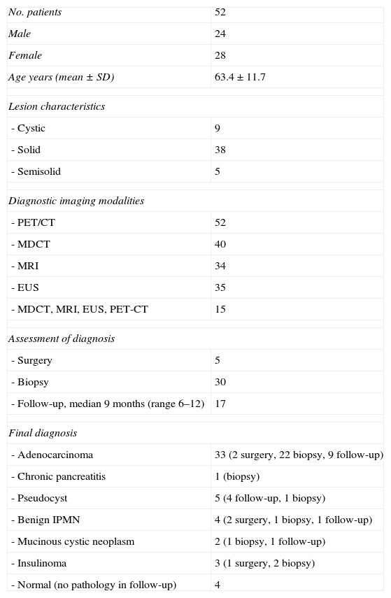
Material and methods

We retrospectively scanned the data of 52 patients who were referred for FDG PET/CT imaging for evaluation of pancreatic lesions greater than 10mm. The diagnostic performances of 4 imaging methods and the impact of PET/CT on the management of pancreatic cancer were defined.
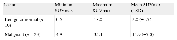
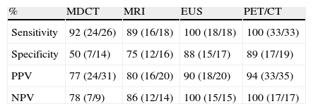
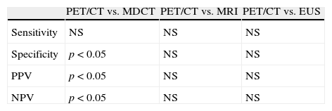
Results

Pancreatic adenocarcinoma was diagnosed in 33 of 52 patients (63%), 15 patients had benign diseases of pancreas (29%), and 4 patients were normal (8%). Sensitivity and NPV of EUS and PET/CT were equal (100%) and higher than MDCT and MRI. Specificity, PPV and NPV of PET/CT were significantly higher than MDCT. However, sensitivities of two imaging methods were not significantly different. There was no significant difference between PET/CT and MRI and EUS for these values. When the cut-off value of SUVmax was 3.2, the most effective sensitivity and specificity values were obtained. PET/CT contributed to the management of pancreatic cancer in 30% of patients.

Conclusion

FDG PET/CT is a valuable imaging method for the diagnosis and management of pancreatic cancer, especially when applied along with EUS as first line diagnostic tools.

Keywords:
18F-Fluorodeoxyglucose Positron Emission Tomography/Computed Tomography
Pancreatic cancer
Diagnosis
Staging
Resumen
Objetivo

El objetivo fue analizar la contribución de la PET/TC con 18F-FDG (FDG PET/TC) en el diagnóstico y tratamiento del cáncer de páncreas en comparación con la tomografía computarizada multidetector (TCMD), la resonancia magnética (RM) y la ecografía endoscópica (EUS).

Material y métodos

Se revisaron retrospectivamente 52 pacientes que fueron remitidos para la evaluación de lesiones pancreáticas mayores de 10mm mediante FDG PET/TC. Se definieron los hallazgos diagnósticos de los 4 métodos de imagen y el impacto de la FDG PET/TC en el tratamiento del cáncer de páncreas.

Resultados

En 33 de los 52 pacientes (63%) se diagnosticó un adenocarcinoma pancreático; 15 pacientes tenían enfermedades benignas del páncreas (29%) y 4 pacientes no mostraron enfermedad pancreática (8%). La sensibilidad y el valor predictivo negativo (VPN) del EUS y la FDG PET/TC fueron iguales (100%) y superior a la TCMD y a la RM. La especificidad, el valor predictivo positivo y el VPN de la FDG PET/TC fueron significativamente mayores que la TCMD; sin embargo, la sensibilidad de 2 métodos de imagen no fue significativamente diferente. No hubo diferencias significativas entre la FDG PET/TC, RM y EUS. Con un punto de corte de SUVmax igual a 3,2 se obtuvieron los valores más efectivos de sensibilidad y de especificidad. La FDG PET/TC contribuyó al manejo clínico del cáncer de páncreas en 30% de los pacientes.

Conclusión

La FDG PET/TC es un método de imagen valioso para el diagnóstico y tratamiento del cáncer de páncreas, especialmente cuando se aplica junto con la EUS como primera línea de herramientas de diagnóstico.

Palabras clave:
Papel de 18F-Fluorodeoxiglucosa por emisión de positrones/tomografía computarizada
Cáncer de páncreas
Diagnóstico
Estadificación

Artículo

Opciones para acceder a los textos completos de la publicación Revista Española de Medicina Nuclear e Imagen Molecular
Socio
Si es usted socio de la Sociedad Española de Medicina Nuclear e Imagen Molecular (SEMNIM) puede acceder al texto completo de los contenidos de la Revista Española de Medicina Nuclear e Imagen Molecular desde los enlaces a la revista publicados en la web de la SEMNIN (enlace a https://semnim.es/iniciar-sesion/), previo inicio de sesión como socio. Si tiene problemas de acceso puede contactar con la Secretaría Técnica de la SEMNIM en el correo electrónico secretaria.tecnica@semnim.es o en el teléfono: + 34 619 594 780
Suscriptor
Suscriptor de la revista

Si ya tiene sus datos de acceso, clique aquí.

Si olvidó su clave de acceso puede recuperarla clicando aquí y seleccionando la opción "He olvidado mi contraseña".
Suscribirse
Suscribirse a:

Revista Española de Medicina Nuclear e Imagen Molecular

Comprar
Comprar acceso al artículo

Comprando el artículo el PDF del mismo podrá ser descargado

Precio 19,34 €

Comprar ahora
Contactar
Teléfono para suscripciones e incidencias
De lunes a viernes de 9h a 18h (GMT+1) excepto los meses de julio y agosto que será de 9 a 15h
Llamadas desde España
932 415 960
Llamadas desde fuera de España
+34 932 415 960
E-mail
Opciones de artículo
Herramientas
es en pt

¿Es usted profesional sanitario apto para prescribir o dispensar medicamentos?

Are you a health professional able to prescribe or dispense drugs?

Você é um profissional de saúde habilitado a prescrever ou dispensar medicamentos

Quizás le interese:
10.1016/j.remn.2023.11.003
No mostrar más