


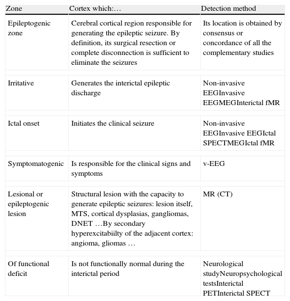
Epilepsy is one of the most frequent chronic neurological disorders, affecting 1–2% of the population. Patients with complex partial drug resistant episodes may benefit from a surgical treatment consisting in the excision of the epileptogenic area. Localization of the epileptogenic area was classically performed with video-EEG and magnetic resonance (MR). Recently, functional neuroimaging studies of nuclear medicine, positron emission tomography (PET) and single photon emission tomography (SPECT) have demonstrated their utility in the localization of the epileptogenic area prior to surgery. Ictal SPECT with brain perfusion tracers show an increase in blood flow in the initial ictal focus, while PET with 18FDG demonstrates a decrease of glucose metabolism in the interictal functional deficit zone.
In this review, the basic principles and methodological characteristics of the SPECT and PET in epilepsy are described. The ictal SPECT injection mechanism, different patterns of perfusion based on the time of ictal, postictal or interictal injection are detailed and the different diagnostic sensitivities of each one of these SPECT are reviewed. Different methods of analysis of the images by subtraction and fusion systems with the MR are described. Similarly, the injection methodology, quantification and evaluation of the images of the PET in epilepsy are described. Finally, the main clinical indications of SPECT and PET in temporal and extratemporal epilepsy are detailed.
La epilepsia es uno de los trastornos neurológicos crónicos más frecuentes, afectando al 1–2% de la población. Los pacientes con crisis parciales complejas resistentes al tratamiento farmacológico pueden beneficiarse de un tratamiento quirúrgico que consiste en la extirpación de la zona epileptógena. Clásicamente la localización de la zona epilpetógena se realiza con vídeo-EEG y resonancia magnética (RM). Recientemente las exploraciones de neuroimagen funcional de medicina nuclear, la tomografía por emisión de positrones (PET) y la tomografía por emisión de fotón único (SPECT) han demostrado utilidad en la localización de la zona epileptógena antes de la cirugía. La SPECT ictal con trazadores de perfusión cerebral demuestra un aumento del flujo sanguíneo en la zona de inicio ictal, mientras que la PET con 18FDG muestra una disminución del metabolismo de la glucosa en la zona de déficit funcional interictal.
En esta revisión se describen los principios básicos y las particularidades metodológicas de la SPECT y la PET en la epilepsia. Se detalla el mecanismo de inyección de la SPECT ictal, los diferentes patrones de perfusión en función del momento de inyección ictal, postictal o interictal y se revisan las diferentes sensibilidades diagnósticas de cada uno de estos SPECT. Se describen diferentes métodos de análisis de las imágenes con sistemas de substracción y fusión con la RM. Del mismo modo, se describe la metodología de inyección, cuantificación y evaluación de las imágenes de la PET en la epilepsia. Finalmente se detallan las principales indicaciones clínicas de la SPECT y de la PET en la epilepsia temporal y extratemporal.
Article
If you experience access problems, you can contact the SEMNIM Technical Secretariat by email at secretaria.tecnica@semnim.es or by phone at +34 619 594 780.

Revista Española de Medicina Nuclear e Imagen Molecular (English Edition)
