


68Ga-PSMA-uptake shows accumulation in the malignant lesions of prostate cancer patients as early as 5min p.i. Studies indicate the value of adding an early image of the pelvis to the imaging protocol of 68Ga-PSMA-11 PET/CT scan showed contradictory results. In this study we planned to assess the significance of an additional early imaging in 68Ga-PSMA I&T PET/CT imaging in prostate cancer patients.
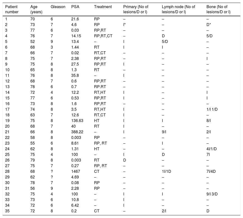
Materials and methodsA total of 35 prostate cancer patients referred to 68Ga-PSMA-I&T PET/CT imaging for restaging of the disease due to suspicion of relapse of the after definitive therapy were enrolled. First an early static pelvis image was obtained at a maximum of 300s following injection of the radiotracer. Sixty minutes post injection a whole-body PET/CT scan was conducted with an emission time of 3min per bed position. The lesions which were categorized as local recurrence, bone lesion and lymph node metastasis in the early images, were compared with the late images in terms of number of lesions detected and SUVmax values.
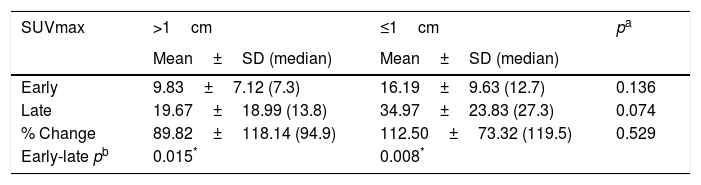
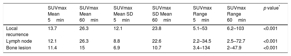
Results68Ga-PSMA-I&T PET/CT was positive in 23 of 35 patients (65.7%). A pathological uptake was observed in the prostatic bed site, in the pelvic lymph nodes, and in the bones in 17 patients (48.5%), 12 patients (34.2%), and 13 patients (37.1%), respectively. In one patient, focal pathological increased uptake in the prostatic bed with a SUVmax value of 5.8 was detected but this lesion disappeared in the late images (Fig. 1). The average SUVmax values of the lesions in the prostatic bed were 13.7±12.1 versus 26.3±23.8 in the 5min and 60min studies respectively (p<0.001). In one patient, the pathological uptake in the lymph node in the early study cleared in the late study, whereas in another accumulation of activity was detected in a pelvic lymph node in the late study, while there was no lymph node detected in the early study. The average SUVmax values of the lymph nodes were 12.1±8.8 versus 26.3±22.6 in the 5min and 60min studies respectively (p<0.001). The average SUVmax values of the bone lesions were 11.4±6.9 versus 15±10.7 in the 5min and 60min studies respectively.
ConclusionOur study is the first in the literature to evaluate the impact of adding an early static pelvic image to the 68GaPSMA-I&T scan, in the detection rate of the lesions. Although there was no marked discordance between the two sets of images, the addition of an early image to the imaging protocol of 68GaPSMA-I&T scan would increase the efficacy of detection of malignant lesions in the pelvis, which might show rapid clearance and has the risk of being masked by the urinary system activity.
El 68Ga-PSMA muestra captación en las lesiones malignas de los pacientes de cáncer de próstata a los 5 minutos postinyección. Los estudios realizados para comprender el valor de añadir una imagen pélvica precoz al protocolo de imagen de 68Ga-PSMA-11 PET/TC muestran resultados contradictorios. En este estudio, planificamos la evaluación de la relevancia de añadir una imagen precoz a 68Ga-PSMA I&T PET/TC en los pacientes con cáncer de próstata.
Materiales y MétodosReunimos a un total de 35 pacientes con cáncer de próstata a quienes se prescribió 68Ga-PSMA-I&T PET/TC para re-estadificar la enfermedad, debido a sospecha de recidiva tras la terapia definitiva. En primer lugar se obtuvo una imagen pélvica estática precoz, a un máximo de 300 segundos tras la inyección del radio-trazador. A los sesenta minutos de la inyección se realizó un PET/TC de cuerpo entero, con un tiempo de emisión de 3min por posición del lecho. Se compararon las lesiones categorizadas en las imágenes precoces como recidiva local, lesión ósea y metástasis ganglionar, con las imágenes tardías en términos de lesiones detectadas y valores SUVmax.
Resultados68Ga-PSMA-I&T PET/TC fue positivo en 23 de los 35 pacientes (65,7%). Se observó una captación patológica en el lecho prostático, en los ganglios pélvicos, y a nivel óseo en 17 pacientes (48,5%), 12 pacientes (34,2%), y 13 pacientes (37,1%), respectivamente. En un paciente, se detectó una captación incrementada patológica focal en el lecho prostático con un valor SUVmax de 5,8, aunque esta lesión desapareció en las imágenes tardías (Figura 1). Los valores SUVmax medios de las lesiones en el lecho prostático fueron 13,7±12,1 frente a 26,3±23,8 en los estudios a los 5min y 60min, respectivamente (p<0,001) En un paciente, la captación patológica ganglionar en el estudio temprano desapareció en el estudio tardío, mientras que en otro paciente la acumulación de actividad se detectó en un ganglio pélvico en el estudio tardío, y no se detectó ningún ganglio en el estudio temprano. Los valores SUVmax ganglionares medios fueron 12,1±8,8 frente a 26,3±22,6 en los estudios a los 5min y 60min, respectivamente (p<0,001). Los valores SUVmax medios de las lesiones óseas fueron 11,4±6,9 frente a 15±10,7 en los estudios a los 5min y 60min, respectivamente.
ConclusiónNuestro estudio es el primero en la literatura que evalúa el impacto de añadir una imagen pélvica estática temprana a 68GaPSMA-I&T, para valorar la tasa de detección de las lesiones. Aunque no se produjo una discordancia marcada entre los dos conjuntos de imágenes, la adición de una imagen temprana al protocolo de imagen de 68GaPSMA-I&T podría incrementar la eficacia en la detección de las lesiones pélvicas malignas, lo cual podría reflejar un rápido aclaramiento, con el riesgo de ser enmascarado por la actividad del sistema urinario.
Article
If you experience access problems, you can contact the SEMNIM Technical Secretariat by email at secretaria.tecnica@semnim.es or by phone at +34 619 594 780.

Revista Española de Medicina Nuclear e Imagen Molecular (English Edition)
