


To determine whether there are significant differences in the apparent diffusion coefficient (ADC) between the apparently normal peritumor white matter surrounding glioblastomas and that surrounding brain metastases.
Material and methodsWe retrospectively reviewed 42 patients with histologically confirmed glioblastomas and 42 patients with a single cerebral metastasis. We measured the signal intensity in the apparently normal peritumor white matter and in the abnormal peritumor white matter on the ADC maps. We used mean ADC values in the contralateral occipital white matter as a reference from which to design normalized ADC indices. We compared mean values between the two tumor types. We calculated the area under the receiver operator characteristic curve and estimated the sensitivity and specificity of the measurements taken.
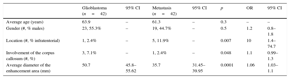
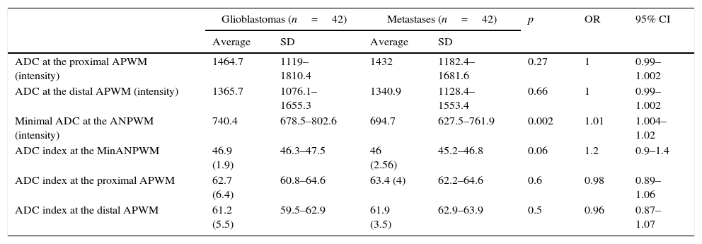
ResultsSupratentorial lesions and compromise of the corpus callosum were more common in patients with glioblastoma than in patients with brain metastases. The maximum diameter of the enhanced area after injection of a contrast agent was greater in the glioblastomas (p<0.001). The minimum ADC value measured in the apparently normal peritumor white matter was higher for the glioblastomas than for the metastases (p=0.002). Significant differences in the ADC index were found only for the minimum ADC value in apparently normal peritumor white matter. The sensitivity and specificity were less than 70% for all variables analyzed.
ConclusionsThere are differences in the ADC values of apparently normal peritumor white matter between glioblastomas and cerebral metastases, but the magnitude of these differences is slight and the application of these differences in clinical practice is still limited.
Encontrar diferencias significativas en la sustancia blanca peritumoral aparentemente normal entre gliobastoma y metástasis cerebral mediante la valoración del coeficiente de difusión aparente (CDA).
Material y métodosSe revisaron retrospectivamente resonancias magnéticas de 42 pacientes con histopatología de glioblastomas y 42 pacientes con metástasis cerebral única. Se realizaron mediciones de intensidad de señal en el mapa de CDA sobre la sustancia blanca peritumoral aparentemente normal (SBPAN) y la sustancia blanca peritumoral alterada (SBPAlt). Se diseñaron índices normalizados de CDA utilizando valores medidos en la sustancia blanca occipital contralateral como referencia. Se compararon las medias para establecer diferencias entre ambos tipos de tumores. Se calculó el área bajo la curva (ROC) y se estimaron la sensibilidad y la especificidad para las mediciones realizadas.
ResultadosLos pacientes con glioblastoma presentaron con mayor frecuencia lesiones supratentoriales y compromiso del cuerpo calloso que los pacientes con metástasis cerebral. El diámetro máximo del área de realce tras la inyección de contraste fue mayor en los glioblastomas (p<0,001). El valor mínimo de CDA medido en la SBPAN fue mayor en los glioblastomas que en las metástasis (p=0,002). Solo se encontraron diferencias significativas en el índice de CDA para el valor mínimo de CDA en la SBPAN. Los valores de sensibilidad y especificidad fueron inferiores al 70% para las variables evaluadas.
ConclusionesExisten diferencias en los valores del CDA de la SBPAN entre glioblastomas y metástasis, pero la magnitud de dicha diferencia es escasa y su aplicación en la práctica clínica aún es limitada.

