


To evaluate whether breast density influences the sensitivity of a computer-assisted detection (CAD) system for the detection of breast cancer.
Material and methodsWe prospectively studied 8750 digital mammograms with an associated CAD system. We used BI-RADS criteria to classify breast density. We calculated the overall sensitivity of the radiologist and of the CAD system, as well as the sensitivity for each projection and type of finding in relation to the mammographic density of the breast. Finally, we analyzed the interval carcinomas. We used SPSS 11 for all statistical analyses.
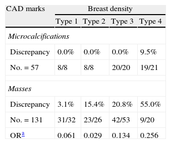
ResultsThe overall sensitivity of the CAD system was 88.5% (95% CI: 83.2–92.7%), and the overall sensitivity of the radiologist was 93.5% (95% CI: 84.4–95.5%). The sensitivity of the craniocaudal view was 81.6% (95% CI: 76.5–90.7%) vs 76.5% (95% CI: 69.3–89.3%) for the mediolateral oblique view. The sensitivity for microcalcifications was 98.6% (95% CI: 96.5–99.7%), and the sensitivity for masses was 83.4% (95% CI: 81.2–91.7%). We detected discrepancies smaller than 20% both for microcalcifications present in the four types of densities and for masses with densities 1 and 2. In masses with density 3 the discrepancy was 20.8% and in those with density 4 it was 55%. The CAD system failed to mark only 9.1% (9/94) of the cancers presenting as masses. Half of the interval carcinomas were found in type 4 density and 75% manifested as masses, asymmetries, and distortions. The CAD system had marked 35.7% of the carcinomas.
ConclusionsThe craniocaudal view was more sensitive, although this difference was not statistically significant. The sensitivity of CAD was high for microcalcifications in all four density types; however, CAD's sensitivity for masses was low in density types 3 and 4. The CAD system only failed to mark 9.1% of the cancers presenting as masses but was not sensitive for the other two radiological findings included in this marking. Half of the interval carcinomas occurred in type 4 densities and 35.7% had been marked by the CAD sysem.
Evaluar si la densidad mamaria influye en la sensibilidad (global y por marcas) para la detección del cáncer de mama de un sistema de detección asistido por ordenador (CAD).
Materiales y métodosEstudio prospectivo de 8.750 mamografías digitales con un sistema CAD asociado. Se clasificaron las densidades mamarias según los criterios BI-RADS. Calculamos la sensibilidad global del radiólogo y del CAD, la sensibilidad por proyección, por hallazgo, en relación con la densidad mamográfica y analizamos los carcinomas de intervalo. Para el análisis estadístico utilizamos el programa SPSS vs 11.
ResultadosSensibilidad global del CAD 88,5% IC del 95% (IC95% 83,2–92,7%), sensibilidad del radiólogo 93,5% IC95% (84,4–95,5%), sensibilidad de la proyección craneocaudal 81,6% IC95% (76,5–90,7%) vs 76,5% IC95% (69,3–89,3%) para oblicuomediolateral, sensibilidad para microcalcificaciones 98,6% IC95% (96,5–99,7%), sensibilidad de marca masa 83,4% IC95% (81,2–91,7%). Detectamos discrepancias menores del 20% tanto para las microcalcificaciones presentes en los 4 tipos de densidades como para las masas con densidades 1 y 2; mientras en las masas con densidad 3 la discrepancia fue 20,8% y en la 4 fue 55%. El CAD solo dejó de marcar el 9,1% (9/94) de los cánceres diagnosticados como masas propiamente dichas. El 50% de los carcinomas de intervalo se produjo en densidad tipo 4 y el 75% se manifestaron como masas, asimetrías y distorsiones. El 35,7% de los carcinomas de intervalo fueron marcados previamente por el CAD.
ConclusionesLa sensibilidad fue mayor en la proyección craneocaudal pero no significativamente. La sensibilidad del CAD fue alta para microcalcificaciones presentes en los 4 tipos de densidades, sin embargo, para la marca masa fue baja en densidades 3 y 4. El CAD sólo dejó de marcar el 9,1% de los cánceres diagnosticados como masas propiamente dichas pero fue muy poco sensible para los otros 2 hallazgos radiológicos incluidos en esta marca. El 50% de los carcinomas de intervalo se produjeron en densidades tipo 4 y el 35,7% fueron marcados previamente por el sistema de detección asistido por ordenador.

