Buscar en
Radiología (English Edition)
Toda la web
Inicio Radiología (English Edition) Diagnostic management of renal colic
Journal Information
Vol. 57. Issue 2.
Pages 113-122 (March - April 2015)
Share
Share
Download PDF
More article options
Visits
2005
Vol. 57. Issue 2.
Pages 113-122 (March - April 2015)
Update in Radiology
Diagnostic management of renal colic
Manejo diagnóstico del cólico renal
Visits
2005
C. Nicolaua,
Corresponding author
cnicolau@clinic.cat

Corresponding author.
, R. Salvadora, J.M. Artigasb
a Servicio de Radiología, Hospital Clínic de Barcelona, Barcelona, Spain
b Servicio de Radiología, Hospital Universitario Miguel Servet, Zaragoza, Spain
This item has received
Article information
Abstract
Full Text
Bibliography
Download PDF
Statistics
Figures (5)
Show moreShow less
Tables (3)
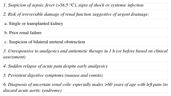
Table 1. Urgent indications for image modalities in patients with acute renal coil.
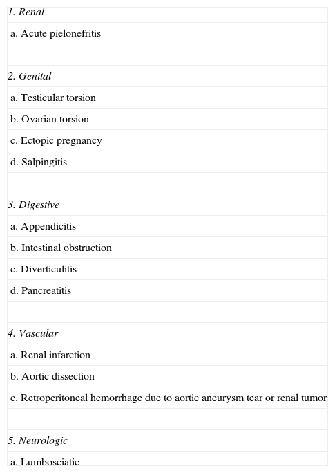
Table 2. Differential diagnosis of reno-ureteral colic.
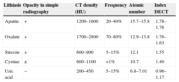
Table 3. Lithiasic composition through DECT.
Show moreShow less
Abstract

Renal colic is a common reason for presentation to emergency departments, and imaging has become fundamental for the diagnosis and clinical management of this condition. Ultrasonography and particularly noncontrast computed tomography have good diagnostic performance in diagnosing renal colic. Radiologic management will depend on the tools available at the center and on the characteristics of the patient. It is essential to use computed tomography techniques that minimize radiation and to use alternatives like ultrasonography in pregnant patients and children. In this article, we review the epidemiology, clinical and radiologic presentations, and clinical management of ureteral lithiasis.

Keywords:
Renal colic
Ultrasonography
Noncontrast computed tomography
Low dose computed tomography
Resumen

El cólico renal es un motivo frecuente de consulta en los Servicios de Urgencias y la imagen diagnóstica se ha convertido en una herramienta fundamental del diagnóstico y manejo clínico. La ecografía y, fundamentalmente, la tomografía computarizada sin contraste permiten diagnosticarlo con un rendimiento elevado. El manejo radiológico va a depender de la disponibilidad del centro y de las características de la población. Es imprescindible usar técnicas de baja dosis de radiación en la tomografía computarizada y técnicas alternativas como la ecografía en embarazadas y niños. En este artículo hacemos una revisión epidemiológica, clínica, radiológica y del manejo clínico de la litiasis ureteral.

Palabras clave:
Cólico renal
Ecografía
Tomografía computarizada sin contraste
Tomografía computarizada baja dosis

Article

These are the options to access the full texts of the publication Radiología (English Edition)
Subscriber
Subscriber

If you already have your login data, please click here .

If you have forgotten your password you can you can recover it by clicking here and selecting the option “I have forgotten my password”
Purchase
Purchase article

Purchasing article the PDF version will be downloaded

Price 19.34 €

Purchase now
Contact
Phone for subscriptions and reporting of errors
From Monday to Friday from 9 a.m. to 6 p.m. (GMT + 1) except for the months of July and August which will be from 9 a.m. to 3 p.m.
Calls from Spain
932 415 960
Calls from outside Spain
+34 932 415 960
E-mail
Article options
Tools
es en pt

¿Es usted profesional sanitario apto para prescribir o dispensar medicamentos?

Are you a health professional able to prescribe or dispense drugs?

Você é um profissional de saúde habilitado a prescrever ou dispensar medicamentos