


To determine the ability of MRI to distinguish between benign and malignant vertebral lesions.
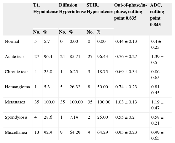
Materials and methodsWe included 85 patients and studied a total of 213 vertebrae (both pathologic and normal). For each vertebra, we determined whether the lesion was hypointense in T1-weighted sequences and whether it was hyperintense in STIR and in diffusion-weighted sequences. We calculated the in-phase/out-of-phase quotient and the apparent diffusion coefficient for each vertebra. We combined parameters from T1-weighted, diffusion-weighted, and STIR sequences to devise a formula to distinguish benign from malignant lesions.
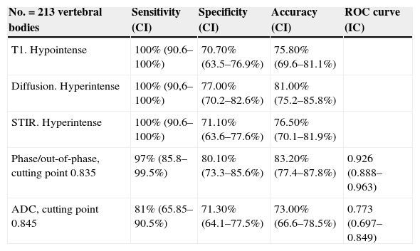
ResultsThe group comprised 60 (70.6%) women and 25 (29.4%) men with a mean age of 67±13.5 years (range, 33–90 y). Of the 85 patients, 26 (30.6%) had a known primary tumor. When the lesion was hypointense on T1-weighted sequences, hyperintense on STIR and diffusion-weighted sequences, and had a signal intensity quotient greater than 0.8, the sensitivity was 97.2%, the specificity was 90%, and the diagnostic accuracy was 91.2%. If the patient had a known primary tumor, these values increased to 97.2%, 99.4%, and 99%, respectively.
ConclusionBenign lesions can be distinguished from malignant lesions if we combine the information from T1-weighted, STIR, and diffusion-weighted sequences together with the in-phase/out-of-phase quotient of the lesion detected in the vertebral body on MRI.
Establecer la capacidad diagnóstica de la RM para distinguir las lesiones vertebrales benignas de las malignas.
Material y métodosIncluimos en el estudio a 85 pacientes con un total de 213 vértebras estudiadas (tanto patológicas como normales). Para cada vértebra determinamos si la lesión era hipointensa en T1 y si era hiperintensa o no en las secuencias STIR y potenciada en difusión. Calculamos el valor del cociente fuera de fase/en fase y el valor del coeficiente de difusión aparente de cada vértebra. A partir de los parámetros T1, difusión y STIR establecimos una combinación diagnóstica de lesión maligna.
ResultadosEl grupo comprendía 60 (70,6%) mujeres y 25 (29,4%) hombres con una edad media de 67±13,5 años (33–90 años). De los 85 pacientes, un total de 26 (30,6%) tenían antecedentes de tumor primario. Cuando la lesión era hipointensa en las imágenes potenciadas en T1, hiperintensa en STIR y en las imágenes potenciadas en difusión, y con un cociente de intensidad de señal mayor de 0,8, la sensibilidad fue del 97,2%; la especificidad del 90% y la exactitud diagnóstica del 91,2%. Si el paciente tenía un tumor primario conocido, los valores se incrementaron hasta el 97,2; 99,4 y 99%, respectivamente.
ConclusiónEs posible distinguir las lesiones benignas de las malignas si valoramos de forma conjunta la señal en T1, STIR y difusión y el cociente fuera de fase/en fase de la lesión detectada con RM en el cuerpo vertebral.

