


Neurological complications are the most frequent type of extracardiac complications of infective endocarditis (IE), and can be the initial manifestation. The objectives of this study were to determine the prevalence of neurological complications in patients with IE and to evaluate whether initial presentation with neurological symptoms causes a diagnostic delay.
Material and methodsWe conducted a retrospective observational study of patients with IE admitted to a tertiary hospital between 2003 and 2020.
ResultsThe study included 222 patients with IE (67% men; mean [SD] age, 66.4 [14.2] years). Neurological complications occurred in 21.2% of patients, with ischaemic stroke (74.5%) and intracerebral haemorrhage (23.4%) being the most frequent. No differences in diagnostic delay were found between the group of patients in whom the disease manifested with neurological complications and the rest of the patients (4.4 vs 4.5; P = .76).
ConclusionsA total of 21.2% of patients with IE presented neurological complications, with ischaemic stroke being the most frequent. Neurological symptoms as the initial manifestation of IE did not lead to a delay in diagnosis.
Las manifestaciones neurológicas son las complicaciones extracardíacas más frecuentes de la endocarditis infecciosa (EI) y pueden ser el síntoma inicial. Los objetivos del estudio fueron determinar la prevalencia de complicaciones neurológicas en los pacientes con EI y evaluar si la clínica neurológica como debut condiciona un retraso diagnóstico.
Material y métodosEstudio de cohortes retrospectivo de pacientes con EI ingresados en un hospital de tercer nivel en el periodo entre 2003 y 2020.
ResultadosSe han revisado 222 pacientes con EI (67% varones, edad 66,4 ± 14,2 años). El 21,2% presentaron complicaciones neurológicas durante el ingreso, siendo el ictus isquémico (74,5%) y la hemorragia cerebral (23,4%), las más frecuentes. No se encontraron diferencias entre el tiempo medio desde el ingreso hasta el diagnóstico en el grupo de pacientes que debutó con focalidad, en comparación con el resto de pacientes (4,4 vs. 4,5; p = 0,76).
ConclusionesUn 21,2% de los pacientes con EI presentaron complicaciones neurológicas durante el ingreso, siendo el ictus isquémico la más frecuente. La clínica neurológica como debut de la EI no conllevó un retraso diagnóstico.
Infective endocarditis (IE) is an acute or subacute, mainly valvular disease of the endocardium, progressing with cardiac and/or non-cardiac systemic symptoms.1 Incidence is estimated at 3–10 cases per 100 000 person-years.2,3 Between 20% and 40% of patients with IE present neurological complications at some point during disease progression, with these complications being the most frequent non-cardiac symptoms.4 Neurological complications can be classified as cerebrovascular (ischaemic stroke, intracranial haemorrhage), infectious (meningitis, brain abscess), and systemic (encephalopathy, seizures).5,6 Ischaemic stroke is the most frequent complication.3,7,8 Therefore, neuroimaging studies are indicated in all patients presenting neurological symptoms.9
Despite advances in the diagnosis and treatment of IE, the associated mortality rate remains high, particularly in patients with neurological complications4,5,10; therefore, multidisciplinary care is essential for its early detection and proper treatment.2,6,9,11–14
This study aims to establish the prevalence of neurological complications in patients with IE, to analyse their clinical and demographic characteristics, and to evaluate whether focal neurological signs as the initial manifestation delays the diagnosis of IE with regard to the patients presenting other systemic and/or cardiac symptoms at onset.
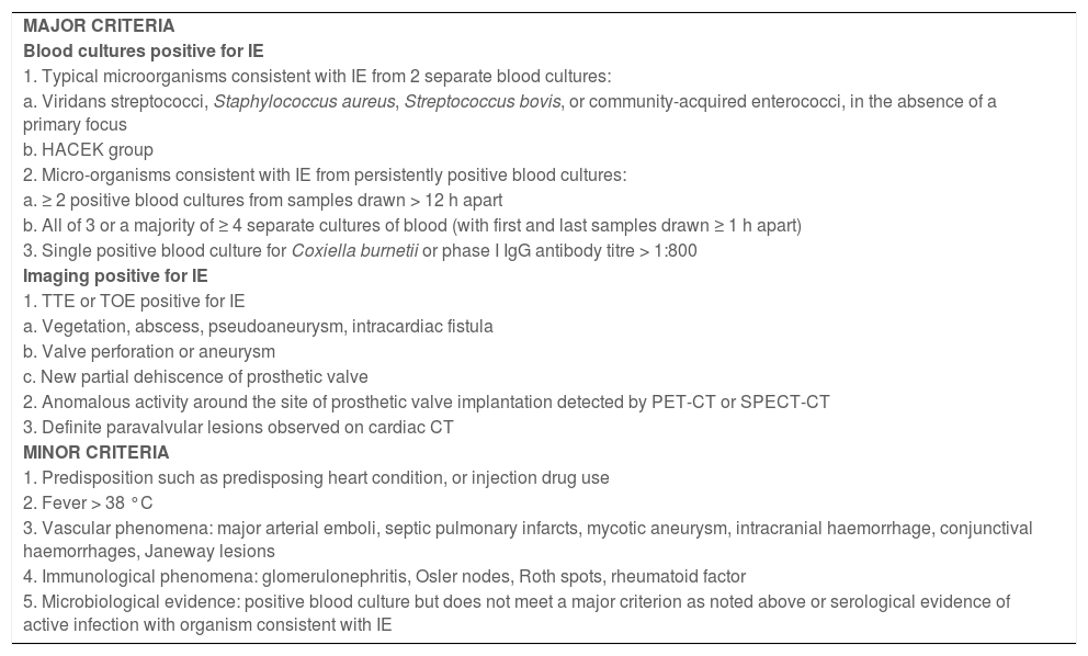
Material and methodsWe conducted an observational, retrospective, analytical cohort study including patients diagnosed with IE at a tertiary-level hospital between January 2003 and December 2020. IE was diagnosed according to the modified Duke criteria for definite IE,15,16 that is, in patients meeting 2 major criteria, one major plus 3 minor criteria, or 5 minor criteria (Table 1). Patients with possible IE were excluded.
Modified Duke criteria.
| MAJOR CRITERIA |
| Blood cultures positive for IE |
| 1. Typical microorganisms consistent with IE from 2 separate blood cultures: |
| a. Viridans streptococci, Staphylococcus aureus, Streptococcus bovis, or community-acquired enterococci, in the absence of a primary focus |
| b. HACEK group |
| 2. Micro-organisms consistent with IE from persistently positive blood cultures: |
| a. ≥ 2 positive blood cultures from samples drawn > 12 h apart |
| b. All of 3 or a majority of ≥ 4 separate cultures of blood (with first and last samples drawn ≥ 1 h apart) |
| 3. Single positive blood culture for Coxiella burnetii or phase I IgG antibody titre > 1:800 |
| Imaging positive for IE |
| 1. TTE or TOE positive for IE |
| a. Vegetation, abscess, pseudoaneurysm, intracardiac fistula |
| b. Valve perforation or aneurysm |
| c. New partial dehiscence of prosthetic valve |
| 2. Anomalous activity around the site of prosthetic valve implantation detected by PET-CT or SPECT-CT |
| 3. Definite paravalvular lesions observed on cardiac CT |
| MINOR CRITERIA |
| 1. Predisposition such as predisposing heart condition, or injection drug use |
| 2. Fever > 38 °C |
| 3. Vascular phenomena: major arterial emboli, septic pulmonary infarcts, mycotic aneurysm, intracranial haemorrhage, conjunctival haemorrhages, Janeway lesions |
| 4. Immunological phenomena: glomerulonephritis, Osler nodes, Roth spots, rheumatoid factor |
| 5. Microbiological evidence: positive blood culture but does not meet a major criterion as noted above or serological evidence of active infection with organism consistent with IE |
Adapted from Habib et al.16
IE was considered nosocomial in patients developing symptoms at least 48 hours after admission to hospital.
Transthoracic echocardiography (TTE) studies were performed in all patients with suspected IE. Subsequently, transoesophageal echocardiography (TOE) was performed in patients with TTE results positive for IE (findings of vegetation, abscess, pseudoaneurysm, perforation, fistula, or valve dehiscence), in those with negative TTE results but strong clinical suspicion of IE, in patients with non-diagnostic TTE findings, and in carriers of prosthetic valves or cardiac devices, as recommended in the diagnostic and therapeutic guidelines of the European Society of Cardiology.16
Blood cultures were performed for all patients with initially suspected IE, according to the protocols of our hospital. For each patient, 3 samples were taken at different times and from different puncture sites, independently of the presence of fever; serology tests were requested for other bacteria (Coxiella burnetii, Mycoplasma, Brucella, Legionella, etc.).
Patients were divided into 2 groups according to whether or not they presented neurological symptoms. Neurological manifestations were classified as follows: ischaemic stroke, intracerebral haemorrhage, seizures, meningitis, and encephalopathy. Ischaemic stroke was diagnosed according to clinical signs and the findings of the head computed tomography (CT) study. A brain magnetic resonance imaging (MRI) study was performed in patients whose CT results did not show acute alterations, or in whom CT findings were not consistent with their clinical symptoms. Haemorrhage was diagnosed according to clinical symptoms and CT findings. No patient underwent an angiography study. Seizures were diagnosed according to the semiology described. Diagnosis of meningitis was based on compatible findings in the cerebrospinal fluid (CSF) analysis. Encephalopathy was defined as impaired level of consciousness without focal neurological signs or neuroimaging anomalies.
The variables analysed in the study are: sex, age at admission, origin of the infection (community-acquired or nosocomial), type of IE (native or prosthetic valve), location of the prosthetic valve, location of the vegetation, microorganism identified, emergency surgery, time from admission to diagnosis, duration of hospital stay, in-hospital mortality, and time to death.
Emergency surgery was defined as the need for a surgical procedure within hours/days, excluding elective surgeries. Indications for emergency surgery were cardiac insufficiency secondary to severe valve involvement, uncontrolled infection (persistent fever with positive blood cultures at 7 days, or development of intracardiac complications), and embolism prevention (vegetations greater than 10 mm).17
The study was approved by the clinical research ethics committee of the region of Aragon (project code PI21/135).
Statistical analysis was conducted using the SPSS statistics software, version 25.0 (IBM Corp.; Armonk, NY, USA). Normally distributed quantitative variables are expressed as means and standard deviations (SD), and were compared with the t test for unpaired samples. Non–normally distributed variables are expressed as medians and the first and third quartiles, and were studied with nonparametric tests. Qualitative variables are expressed as percentages, and were compared using the chi-square test. Values of P < .05 were considered statistically significant.
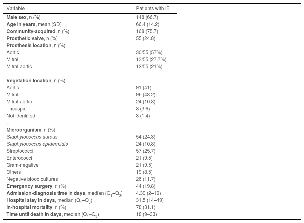
ResultsOur sample included a total of 222 patients diagnosed with definite IE (67% men; mean [SD] age of 66.4 [14.2] years). TTE and/or TOE were performed in 221 patients (99.5%); blood culture results were positive in 196 patients (88.2%). Regarding the modified Duke criteria for IE, 192 patients (86.5%) met 2 major criteria, and the remaining 30 (13.5%) met one major and 3 minor criteria. Demographic, clinical, and microbiological variables are shown in Table 2.
Demographic, clinical, and microbiological variables in patients with infective endocarditis (n = 222).
| Variable | Patients with IE |
|---|---|
| Male sex, n (%) | 148 (66.7) |
| Age in years, mean (SD) | 66.4 (14.2) |
| Community-acquired, n (%) | 168 (75.7) |
| Prosthetic valve, n (%) | 55 (24.8) |
| Prosthesis location, n (%) | |
| Aortic | 30/55 (57%) |
| Mitral | 13/55 (27.7%) |
| Mitral-aortic | 12/55 (21%) |
| – | |
| Vegetation location, n (%) | |
| Aortic | 91 (41) |
| Mitral | 96 (43.2) |
| Mitral-aortic | 24 (10.8) |
| Tricuspid | 8 (3.6) |
| Not identified | 3 (1.4) |
| – | |
| Microorganism, n (%) | |
| Staphylococcus aureus | 54 (24.3) |
| Staphylococcus epidermidis | 24 (10.8) |
| Streptococci | 57 (25.7) |
| Enterococci | 21 (9.5) |
| Gram-negative | 21 (9.5) |
| Others | 19 (8.5) |
| Negative blood cultures | 26 (11.7) |
| Emergency surgery, n (%) | 44 (19.8) |
| Admission-diagnosis time in days, median (Q1–Q3) | 4.39 (2–10) |
| Hospital stay in days, median (Q1–Q3) | 31.5 (14–49) |
| In-hospital mortality, n (%) | 78 (31.1) |
| Time until death in days, median (Q1–Q3) | 18 (9–33) |
IE: infective endocarditis; SD: standard deviation.
Forty-seven patients (21.2%) presented neurological symptoms either at onset or as a complication during disease progression. Of these, 12 presented more than one complication. Neurological symptoms were the initial manifestation of IE in 16 patients (8% of the total sample; 34% of patients with these complications). The most frequent neurological symptoms at onset were ischaemic stroke (15/16 patients [93.7%]) and meningitis (2/16 [12.5%]).
The most frequent neurological complication among all patients with IE was ischaemic stroke (35/47 [74.5%]). Among these patients, ischaemic lesions involved the carotid territory in 22/35 (62.8%), the vertebrobasilar territory in 5/35 (14.3%), and both territories in 8/35 (29.9%). Ischaemic lesions affected both hemispheres in 12/35 patients (34.3%). Fifteen of these 35 patients (42.8%) presented 2 or more lesions in multiple territories. Haemorrhagic transformation occurred in 3/35 (8.8%).
Cerebral haemorrhage was a complication in 11/47 patients (23.4%), with lobar intraparenchymal haemorrhage being the most frequent type (5/11 patients [45.5%]). Lobar, deep, and subarachnoid haemorrhages were observed simultaneously in 3/11 patients (27.3%).
Seven of the 47 patients (14.9%) presented seizures; only one presented status epilepticus, which progressed favourably.
Bacterial meningitis was confirmed in 6/47 patients (12.8%). The least frequent complication was encephalopathy, which was observed in a single patient (2.1%).
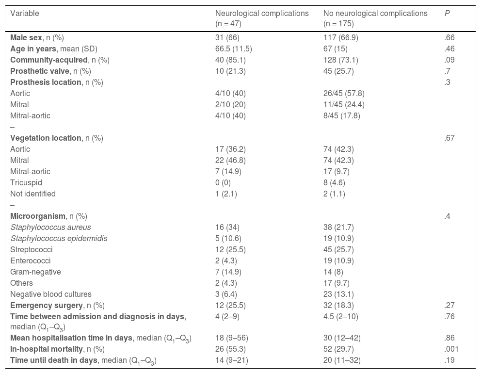
Comparison of data between the 2 subgroups (Table 3) revealed a higher mortality rate in the group of patients with neurological complications (55.3%, vs 29.7% in patients without these complications; P < .01). This group also presented higher prevalence of Staphylococcus aureus (34% vs 21.7%) and mitral or mitral-aortic involvement (61.7% vs 52%), and shorter hospital stays (18 vs 30 days); these differences were not statistically significant.
Demographic, clinical, and microbiological variables in patients with infective endocarditis, by subgroup.
| Variable | Neurological complications (n = 47) | No neurological complications (n = 175) | P |
|---|---|---|---|
| Male sex, n (%) | 31 (66) | 117 (66.9) | .66 |
| Age in years, mean (SD) | 66.5 (11.5) | 67 (15) | .46 |
| Community-acquired, n (%) | 40 (85.1) | 128 (73.1) | .09 |
| Prosthetic valve, n (%) | 10 (21.3) | 45 (25.7) | .7 |
| Prosthesis location, n (%) | .3 | ||
| Aortic | 4/10 (40) | 26/45 (57.8) | |
| Mitral | 2/10 (20) | 11/45 (24.4) | |
| Mitral-aortic | 4/10 (40) | 8/45 (17.8) | |
| – | |||
| Vegetation location, n (%) | .67 | ||
| Aortic | 17 (36.2) | 74 (42.3) | |
| Mitral | 22 (46.8) | 74 (42.3) | |
| Mitral-aortic | 7 (14.9) | 17 (9.7) | |
| Tricuspid | 0 (0) | 8 (4.6) | |
| Not identified | 1 (2.1) | 2 (1.1) | |
| – | |||
| Microorganism, n (%) | .4 | ||
| Staphylococcus aureus | 16 (34) | 38 (21.7) | |
| Staphylococcus epidermidis | 5 (10.6) | 19 (10.9) | |
| Streptococci | 12 (25.5) | 45 (25.7) | |
| Enterococci | 2 (4.3) | 19 (10.9) | |
| Gram-negative | 7 (14.9) | 14 (8) | |
| Others | 2 (4.3) | 17 (9.7) | |
| Negative blood cultures | 3 (6.4) | 23 (13.1) | |
| Emergency surgery, n (%) | 12 (25.5) | 32 (18.3) | .27 |
| Time between admission and diagnosis in days, median (Q1–Q3) | 4 (2–9) | 4.5 (2–10) | .76 |
| Mean hospitalisation time in days, median (Q1–Q3) | 18 (9–56) | 30 (12–42) | .86 |
| In-hospital mortality, n (%) | 26 (55.3) | 52 (29.7) | .001 |
| Time until death in days, median (Q1–Q3) | 14 (9–21) | 20 (11–32) | .19 |
SD: standard deviation.
No statistically significant differences were observed in the median time between admission and diagnosis between patients with neurological symptoms at onset and the remaining patients (4 vs 4.5 days; P = .076).
DiscussionThe aims of this study were to establish the prevalence of neurological complications in IE and to evaluate whether diagnosis is delayed in patients presenting these complications at onset, compared to the remaining patients.
The diagnosis of IE is based on clinical signs, echocardiography findings, and microbiology study results, in accordance with the modified Duke criteria,15,16 and can be a challenge in patients who initially present with symptoms secondary to complications.12
The main pathogenic mechanism of neurological complications, whether symptomatic or subclinical, is embolisation of fragments from the vegetations,6 together with the systemic inflammatory response to infection and small-vessel vasculitis in the central nervous system (CNS).18 The main risk factors for embolisation are delayed onset of antibiotic treatment, vegetations larger than 10 mm or involving valves on the left side of the heart, and positive blood culture results for S. aureus or Streptococcus bovis.6,13,14
The study population included predominantly men in the sixth decade of life, with mainly mitral and aortic valve involvement; 20% were prosthetic valve carriers, and microbiology study results suggested a similar distribution of infections to those reported in other studies of IE.2,5
Neurological complications were observed in 21.1% of patients; this is similar to the rates reported in other studies, which range from 20% to 40%4,5,7 This patient subgroup presented slightly higher prevalence of mitral valve involvement, as reported by other authors, although this difference was not significant. Our analysis distinguished isolated mitral valve involvement from mitral-aortic involvement; if these groups are combined, the difference is greater (61.7% vs 52%).
Regarding aetiology, S. aureus infection is frequently reported. This was also observed in our cohort, though with no significant difference between groups (34% vs 21.7%).8
The in-hospital mortality rate in our cohort (31.1%) is at the upper bound of the rates reported by other groups (15%–30%), and similar to that described by Marques et al.19 This may be due to the percentage of patients undergoing cardiac surgery (associated with better prognosis), which was lower than that described in the literature (40%–50%),2,4,8,10 although a slightly higher percentage of patients with neurological complications underwent surgery (25.5% vs 18.3%). It may also partially be explained by the lack of a cardiac surgery department at our hospital, which may have hindered multidisciplinary assessment, and by the potential presence of comorbidities in these patients, which are not analysed due to the retrospective nature of the study.
As observed in our results, the development of neurological complications is associated with poorer prognosis, contributing to a significant increase in in-hospital mortality compared to patients without these complications (55% vs 30%).3,5,7,11 In addition, patients with neurological complications presented shorter hospital stays (18 vs 30 days); however, this difference was not statistically significant. This finding may have been influenced by the higher mortality rate in these patients in the first 2 weeks after admission.
It should be noted that 8% of all patients with IE, and 34% of those with at least one neurological complication, presented neurological symptoms as the initial manifestation of IE, with no signs of concomitant infection; these percentages are similar to those reported by Castilla-Guerra et al.11 (10%–15%). In our study, neurological symptoms as the initial manifestation were not associated with a delay in diagnosis with respect to patients presenting systemic and infectious symptoms at onset.
Ischaemic stroke was the most frequent neurological complication, observed in 74.5% of these patients; this is similar to the rate reported by Silver et al.3 (70%) and higher than that described by García-Cabrera et al.4 (56%). Forty-three percent of patients presented 2 or more ischaemic lesions in different vascular territories and at different stages of progression, as has been reported by other authors.5,8 This neuroimaging finding may be suggestive of IE, particularly in patients with history of valve involvement and/or with fever. An association with S. aureus infection, mitral valve involvement, and vegetation size has also been reported.5,13
Haemorrhage was the second most frequent complication (23.4%), and was associated with a high mortality rate (54.5%); this is consistent with the data presented by Dourado Sotero et al.5 These haemorrhages often result from haemorrhagic transformation of ischaemic stroke, although only 3 cases were observed in our study. They may also occur in patients with mycotic aneurysms or septic necrotic arteritis with small-vessel rupture.5,12 We were unable to analyse the presence of mycotic aneurysms, as no angiography studies were performed in the study cohort; these lesions were not described in the patient undergoing MR angiography.
We observed a higher percentage of patients with meningitis (12.8%) than that reported in other studies.4,5 In half of cases, the aetiological agent was S. aureus. The pathogenic mechanism may have been the development of microabscesses in the brain. Two-thirds of patients presented CSF findings compatible with aseptic meningitis, as described in the literature (discrete mononuclear pleocytosis),6,13 with one-third presenting cytobiochemistry results compatible with acute bacterial meningitis, with pleocytosis, low glucose levels, and elevated protein levels.
We observed lower prevalence of encephalopathy than other authors5; this complication was probably underdiagnosed due to the non-specificity of its symptoms in the context of severe infection.
The strengths of the study include the long study period and the large sample size. However, it also presents some limitations. Due to the observational, retrospective design of the study, some data were missing for patients diagnosed prior to the implementation of electronic record-keeping. Furthermore, due to the inability to precisely determine the progression time of the symptoms that led patients to visit the emergency department, we were forced to study the time from admission to diagnosis. Finally, we may have underestimated the prevalence of ischaemic lesions due to the fact that some patients were studied with head CT only; MRI is a more sensitive imaging technique for detecting embolisms in the CNS,12,13 even in cases in which neurological examination yields normal findings.20
There is a need for prospective studies analysing the presence of silent embolism and seeking to determine what factors may influence prognosis and mortality.
ConclusionsIn this series, 21.2% of patients with IE presented neurological complications, with ischaemic stroke and intraparenchymal haemorrhage being the most frequent. Presence of neurological symptoms as the initial manifestation of IE was not associated with delays in diagnosis compared to the remaining patients. However, given the high morbidity and mortality rates associated with IE, it should be considered as an aetiological diagnosis in patients presenting acute neurological symptoms with some atypical finding, particularly due to the implications of this diagnosis in early therapeutic management.
FundingThis study received no funding of any kind.
Conflicts of interestThe authors have no conflicts of interest to declare.
