Buscar en
Medicina Clínica Práctica
Toda la web
Inicio Medicina Clínica Práctica Round pneumonia in an elderly woman
Información de la revista
Vol. 3. Núm. 6.
(Noviembre - Diciembre 2020)
Compartir
Compartir
Descargar PDF
Más opciones de artículo
Vol. 3. Núm. 6.
(Noviembre - Diciembre 2020)
Images in medicine
Open Access
Round pneumonia in an elderly woman
Neumonía redonda en mujer de edad avanzada
Visitas
1573
Javier Navarro-Estevaa,
Autor para correspondencia
jnesteva7@hotmail.com

Corresponding author.
, Juan María Arroyo-Delgadob, Federico López-Sagastumeb
a Neumología, Hospital San Roque Maspalomas, Maspalomas, San Bartolomé de Tirajana, Spain
b Medicina Interna, Hospital San Roque Maspalomas, Maspalomas, San Bartolomé de Tirajana, Spain
Este artículo ha recibido

Under a Creative Commons license
Información del artículo
Texto completo
Bibliografía
Descargar PDF
Estadísticas
Figuras (4)
Mostrar másMostrar menos
Texto completo

In adults, round pneumonias may be due to infections like Q fever, legionella micdadei, streptococcus pneumoniae, hemophilus influenzae, or rickettsia typhi, and noninfectious causes like bronchogenic carcinoma, round atelectasis, organizing pneumonia, bronchopulmonary sequestration, granulomatosis with polyangiitis, septic pulmonary emboli, rheumatoid nodules or sarcoidosis. They are rarely seen in adults because the development of the pores of Kohn and canals of Lambert is completed by the age of eight.1,2 A 70 year-old woman exsmoker with coronary heart disease and diabetes was admitted with high fever, purulent sputum, dyspnea and anorexia. She was tachypneic, tachycardic and required a FiO2 of 50% to ensure adequate oxygenation. Inflammatory markers were markedly elevated – leucocytes, neutrophils, procalcitonin. A presumptive diagnosis of community acquired pneumonia was made, CURB65: 4/5.

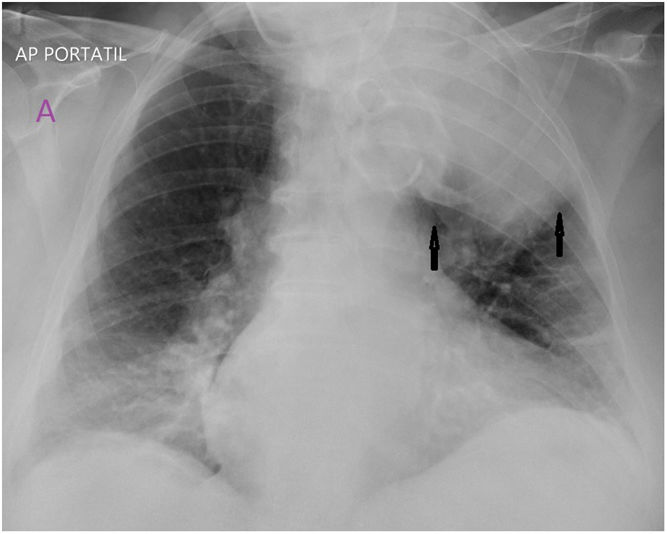
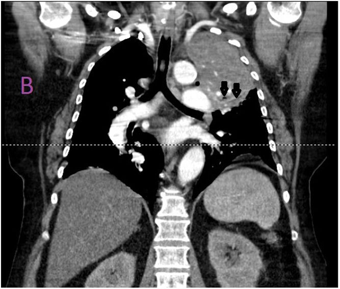
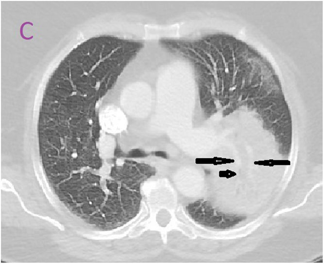
Round left upper lobe infiltrate with clear borders (arrows) is shown in Fig. A. The CT identifies a homogeneous mass-like infiltrate with some air bronchogram (short arrows) and no distorsion of the pulmonary vessels (long arrows) (Figs. B, C). On the day of discharge, the “mass” is vanished (Fig. D).

Figure A
(0,03MB).
Figure B
(0,06MB).
Figure C
(0,04MB).
Figure D
(0,04MB).

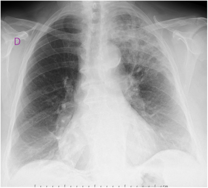
The blood cultures were positive for steptococcus pneumoniae. The response to antibiotics was adequate and the patient was discharged ten days later. The dissapearance of the infiltrate may help avoid an unneccessary bronchoscopy.

References
[1]
B.A. Cunha, A. Gran, J. Simon.
Round pneumonia in a 50 year-old man.
Respir Care, 58 (2013), pp. e80-e82
[2]
J.J. Camargo, S.M. Camargo, T.N. Machuca, F.A. Perin.
Round pneumonia: a rare condition mimicking bronchogenic carcinoma. Case report and review of the literature.
Sao Paulo Med J, 126 (2008), pp. 236-238
Copyright © 2020. The Author(s)
Opciones de artículo
Herramientas
es en pt

¿Es usted profesional sanitario apto para prescribir o dispensar medicamentos?

Are you a health professional able to prescribe or dispense drugs?

Você é um profissional de saúde habilitado a prescrever ou dispensar medicamentos

Quizás le interese:
10.1016/j.mcpsp.2022.100360
No mostrar más