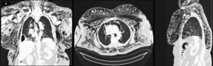
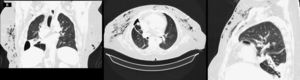
A 73-year-old woman, with a history of arterial hypertension, type 2 diabetes mellitus and obesity. She went to the Emergency Department for dyspnea, having been hospitalized with the diagnosis of decompensated heart failure with type 2 respiratory failure. During hospitalization, for the etiological study of a pleural effusion, a diagnostic thoracentesis was performed, where an empyema was objectified and a chest drain was placed and underwent fibrinolytic therapy and DNAse, having also completed antibiotic therapy with ceftriaxone and azithromycin. As an intercurrence of the chest tube placement, the patient developed grade V subcutaneous emphysema. Thoracic- abdominal-pelvic computed tomography (CT) scan revealed "exuberant bilateral cervical-thoraco- abdominal subcutaneous emphysema with inferior extension to the left and right iliac fossa reaching the inguinal region and exuberant mediastinal emphysema" (Image A). Two subcutaneous drains were placed in the anterior thoracic region along the 3rd intercostal space with significant improvement and almost complete resolution of emphysema in 2 weeks (Image B).
Citescore 2024
0,7
SJR
SJR es una prestigiosa métrica basada en la idea de que todas las citaciones no son iguales. SJR usa un algoritmo similar al page rank de Google; es una medida cuantitativa y cualitativa al impacto de una publicación.
Ver másSJR 2024
0,140
SNIP
SNIP permite comparar el impacto de revistas de diferentes campos temáticos, corrigiendo las diferencias en la probabilidad de ser citado que existe entre revistas de distintas materias.
Ver másSNIP 2024
0,282
Porcentaje de aceptación
La tasa de aceptación de las revistas se calcula dividiendo el número total de artículos aceptados (inicialmente y después de la revisión estándar) por el número total de envíos (aceptados y rechazados).
Ver másPorcentaje de aceptación
63 %
Ver más métricas