
metricas
Enfermedades Infecciosas y Microbiología Clínica Dengue warning signs persisting post-48h demand monitoring to avert severe compl...

Información de la revista
Vol. 44. Núm. 1.
(Enero 2026)
Cita
Cita
Compartir
Descargar PDF
Más opciones de artículo
Visitas
832
Vol. 44. Núm. 1.
(Enero 2026)
Original article
Acceso a texto completo

Dengue warning signs persisting post-48h demand monitoring to avert severe complications

Los signos de alerta de dengue que continúan después de 48 horas necesitan seguimiento para prevenir complicaciones severas
Visitas
832
Verónica Andrade-Almaráz,, José Pedro Elizalde-Díaz
Autor para correspondencia
pedroelizaldediaz@gmail.com

Corresponding author.
Research Service, Hospital Regional de Alta Especialidad “Centenario de la Revolución Mexicana”, Instituto de Seguridad y Servicios Sociales de los Trabajadores del Estado, Emiliano Zapata, Morelos, Mexico
Este artículo ha recibido
Información del artículo
Resumen
Texto completo
Bibliografía
Descargar PDF
Estadísticas
Figuras (2)
fig0005
fig0010
Tablas (4)
Table 1. Gender-based differences in age and symptom duration among dengue patients with and without warning signs.
Tablas
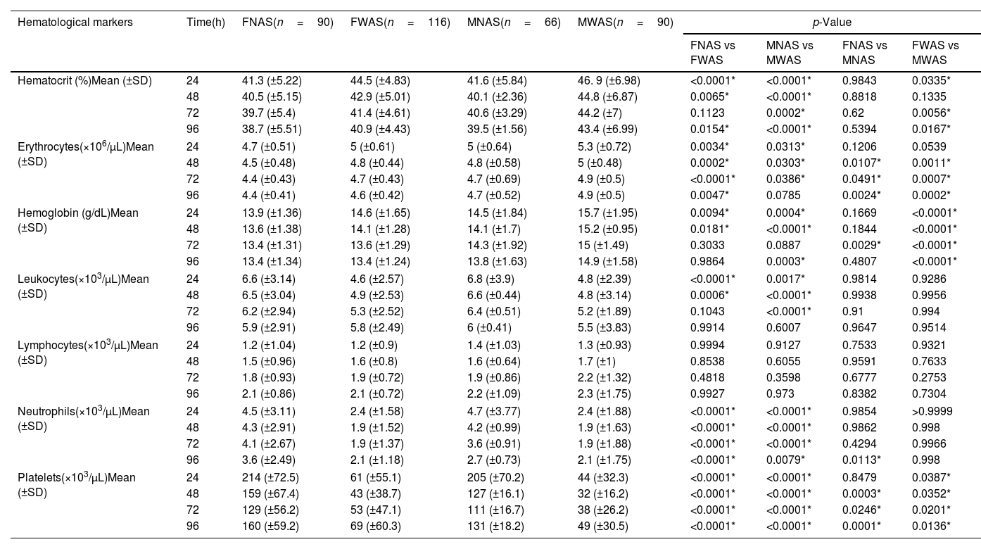
Table 2. Variations according to gender and time in hematological markers between patients with dengue with and without warning signs.
Tablas
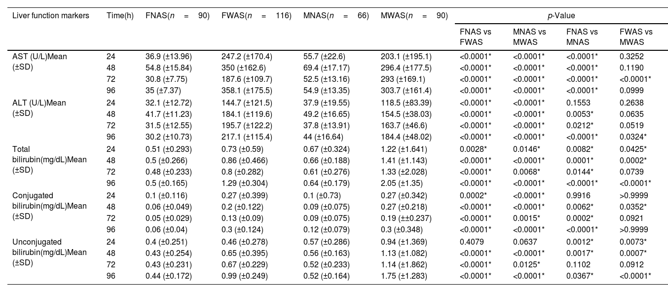
Table 3. Hepatic markers in dengue: differential patterns by gender, time and clinical severity.
Tablas
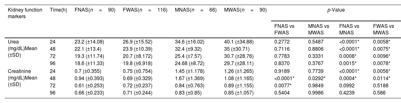
Table 4. Time and sex-specific kidney function profiles in dengue.
Tablas
Material adicional (1)
Abstract
Introduction

Dengue virus infection remains a major public health challenge in tropical regions, with heterogeneous progression to severe disease.

Methods

We conducted a prospective longitudinal study of 362 laboratory-confirmed dengue patients, stratified by sex and the presence of warning signs. Serial hematological, hepatic and renal markers were collected over 96h (at 24h, 48h, 72h, and 96h after symptom onset) to analyze their dynamic evolution.

Results

Patients with warning signs persisting beyond 48h exhibited progressive hematological and hepatic deterioration, unlike those without warning signs. This was characterized by progressively declining platelet and neutrophil counts, elevated hematocrit, and rising AST/ALT levels. These worsening trends were more pronounced in women, who also showed a higher prevalence of warning signs (32% vs 25% in men).

Conclusion

The persistence of warning signs beyond 48h represents a clinically relevant threshold for hematological and hepatic deterioration, supporting the need for protocolized monitoring – including serial platelet counts, hematocrit, and liver enzymes – during this critical window to enable early intervention in high-risk patients.

Keywords:
Dengue
Warning signs
Hematological disturbances
Hepatic dysfunction
Longitudinal
Resumen
Introducción

La infección por el virus del dengue representa un importante reto de salud pública en regiones tropicales, con una progresión heterogénea hacia formas graves.

Métodos

Se realizó un estudio longitudinal prospectivo en 362 pacientes con dengue confirmado por laboratorio, estratificados por sexo y presencia de signos de alarma. Se recolectaron marcadores hematológicos, hepáticos y renales de forma seriada durante 96 horas (a las 24, 48, 72 y 96 horas tras el inicio de síntomas) para analizar su evolución dinámica.

Resultados

Los pacientes con signos de alarma persistentes más allá de 48 horas exhibieron un deterioro hematológico y hepático progresivo, a diferencia de aquellos sin signos de alarma. Esto se caracterizó por descensos progresivos en el recuento de plaquetas y neutrófilos, hematocrito elevado y niveles aumentados de AST/ALT. Estas tendencias de empeoramiento fueron más pronunciadas en mujeres, quienes también mostraron una mayor prevalencia de signos de alarma (32% vs. 25% en hombres).

Conclusión

La persistencia de signos de alarma más allá de 48 horas representa un umbral clínicamente relevante de deterioro hematológico y hepático, lo que respalda la necesidad de un monitoreo protocolizado—que incluya recuentos seriados de plaquetas, hematocrito y enzimas hepáticas—durante esta ventana crítica para permitir una intervención temprana en pacientes de alto riesgo.

Palabras clave:
Dengue
Signos de alarma
Alteraciones hematológicas
Disfunción hepática
Longitudinal
Texto completo
Introduction

Dengue, an arboviral disease affecting millions of people worldwide, poses a significant public health challenge, particularly in tropical and subtropical regions such as Latin America.1 The primary transmission vectors are mosquitoes of the genus Aedes, particularly Aedes aegypti. The incidence of this arbovirus has increased alarmingly due to factors such as urbanization, climate change, and population mobility.2

According to the World Health Organization (WHO), in 2024, 112 countries reported a total of 14.3 million dengue cases, resulting in 10,576 deaths.3 In Latin America, the Pan American Health Organization (PAHO) has documented recurrent outbreaks with considerable health and economic impacts, driven by costs associated with medical care, hospitalizations, and lost productivity.4 For example, in Mexico, the Ministry of Health reported 54,406 confirmed cases in 2023, while by 2024 the number increased to 125,160, highlighting the growing threat of dengue in the region.5

The dengue virus (DENV) is a member of the Flaviviridae family and includes four distinct serotypes (DENV-1 to DENV-4), each with unique antigenic properties.6 The clinical presentation of dengue varies widely, from asymptomatic infections to severe cases involving hemorrhage, plasma leakage, shock, and multi-organ failure.7,8 To standardize diagnosis and treatment, the World Health Organization (WHO) established the 2009 Guidelines for Diagnosis, Treatment, Prevention, and Control of Dengue, which classifies the disease into three categories: dengue without warning signs, dengue with warning signs, and severe dengue.9

Dengue without warning signs is defined by acute-onset high fever (38–40°C) accompanied by at least two of the following: severe headache, retro-orbital pain, myalgia, arthralgia, nausea, vomiting, maculopapular rash, or leukopenia, with occasional positive tourniquet test results. This form typically lasts 2–7 days and is managed with outpatient care, including rest, oral rehydration, and analgesics, as it rarely progresses to systemic complications during the febrile phase.9

Dengue with warning signs includes the above symptoms along with clinical indicators of disease progression, which appear during the critical phase (days 3–5) as fever subsides. These warning signs consist of severe abdominal pain, persistent vomiting, fluid accumulation (e.g., ascites or pleural effusion), mucosal bleeding (e.g., epistaxis or gingival bleeding), lethargy or irritability, and hepatomegaly exceeding 2cm below the costal margin. These findings suggest plasma leakage or early organ dysfunction, requiring hospitalization and intravenous fluid therapy.9,10

Severe dengue involves life-threatening complications such as shock from severe plasma leakage (dengue shock syndrome), characterized by tachycardia, weak pulse, cold extremities, and delayed capillary refill; significant hemorrhage (e.g., hematemesis or melena) leading to hemodynamic instability; or severe organ impairment, including acute hepatitis (AST/ALT >1000U/L), myocarditis, encephalitis, or renal failure. Such cases demand urgent intensive care, with aggressive fluid resuscitation, blood product transfusions if needed, and organ support measures.9–11

Hematological markers, including thrombocytopenia, hemoconcentration, and leukopenia, along with elevated hepatic enzymes (AST and ALT), serve as well-established indicators of disease severity, reflecting the virus's effects on the hematopoietic system and liver.12 In contrast, the clinical significance of renal involvement in severe dengue remains unclear due to conflicting evidence; proposed mechanisms include direct tubular injury or hypoperfusion secondary to shock.13

The clinical management of dengue presents significant challenges due to the disease's complexity. Although diagnostic and prognostic tools have improved, early identification of patients at risk for severe progression remains difficult for clinicians.14 While recent studies highlight the predictive value of warning signs, uncertainties persist regarding the optimal timing for intensive monitoring and intervention, limiting the effectiveness of current management strategies.15 A multicenter study in endemic areas demonstrated that prolonged symptom duration correlates with increased risk of hematological, hepatic, and systemic complications, underscoring the need for well-defined temporal thresholds to improve clinical outcomes.8

This study hypothesized that extended clinical monitoring (>48h) is critical for dengue patients with warning signs due to progressive hematological and hepatic dysfunction. To address the lack of evidence-based thresholds for surveillance, we conducted a sex-stratified analysis of 362 laboratory-confirmed cases, evaluating temporal patterns in hematocrit, platelet counts, and liver enzymes. Our findings confirm that warning signs persisting beyond 48h predict severe complications, mandating protocolized monitoring to improve outcomes.

Materials and methods

This study employed a prospective longitudinal design to evaluate the temporal evolution of clinical and laboratory markers in patients with confirmed dengue. Follow-up was conducted at the General Hospital of Emiliano Zapata, Morelos, Mexico, during the epidemic periods from 2019 to 2021. We selected this design because it allowed for the analysis of intraindividual changes in the parameters of interest over time, specifically during the critical phase of the disease. Standardized evaluation points were established at 24, 48, 72, and 96h after symptom onset, enabling us to capture the temporal dynamics of hematological, hepatic, and renal markers in relation to the presence of warning signs.

Participants and selection criteria

The study included 362 patients with laboratory-confirmed dengue (PCR or serology). Inclusion criteria were1: age over 18 years,2 clinical presentation compatible with dengue according to WHO 2009 criteria, and3 laboratory confirmation. Cases involving pregnancy, coinfection with other arboviruses, or decompensated chronic diseases were excluded. The final sample was stratified into four groups based on sex and the presence of warning signs: women without warning signs (FNAS, n=90), women with warning signs (FWAS, n=116), men without warning signs (MNAS, n=66), and men with warning signs (MWAS, n=90). This stratification allowed for the analysis of differences in disease progression according to these key variables.

Sample and data collection procedures

A standardized protocol was implemented for data collection, which included demographic characteristics and detailed clinical information. Clinical parameters included fever duration, rash, myalgia, headache, and other symptoms associated with dengue. A blood sample was taken from each participant at 24, 48, 72, and 96h for analysis in the clinical laboratory. Laboratory data included hematological parameters (platelet count, white blood cell count, and hematocrit), markers of liver damage (aspartate aminotransferase, alanine aminotransferase, total bilirubin, conjugated bilirubin, and unconjugated bilirubin), and markers of kidney damage (urea and creatinine). All data were recorded prospectively to minimize recall bias and ensure accuracy.

Dengue classification criteria

Disease severity was assessed using the WHO 2009 dengue classification guidelines. Patients were categorized as having either dengue without warning signs or dengue with warning signs (e.g., abdominal pain, persistent vomiting, mucosal bleeding).

Differential diagnosis by laboratory tests

Serological testing was performed using the Panbio® Dengue Early ELISA for NS1 antigen detection and the Panbio® Dengue IgM Capture ELISA for IgM antibody detection, following manufacturer protocols.

For molecular confirmation, viral RNA was extracted from serum samples using the QIAamp® Viral RNA Mini Kit. Real-time RT-PCR amplification was carried out with the VIASURE Multiplex Zika, Dengue y Chikungunya on a CFX96 Touch™ Real-Time PCR. This system allows for the specific detection and differentiation of Zika, Dengue, and/or Chikungunya viruses; however, it does not differentiate between virus serotypes. Each cycle included appropriate positive and negative controls, with cycle thresholds ≤40 considered positive.

Statistical analysis

A comprehensive statistical approach was implemented using GraphPad Prism 9 (version 8.0.2). Initially, a descriptive analysis of all variables was performed, calculating measures of central tendency and dispersion (means, standard deviations) to characterize the study population. Subsequently, mixed-effects models were applied to evaluate temporal changes in hematological, hepatic, and renal markers, specifically accounting for intra-subject correlation in serial measurements. These models included fixed effects for study group (FNAS, FWAS, MNAS, MWAS), evaluation time (24h, 48h, 72h, 96h), and their interaction, while random effects were incorporated to capture interindividual variability.

For multiple comparisons between groups and time points, Tukey's test was applied as a post-hoc method, which controls the type I error rate in analyses involving multiple comparisons. This approach was particularly relevant for identifying specific differences in the temporal evolution of parameters among patient subgroups. All analyses were performed with a significance level of *p<0.05. The a priori sample size calculation indicated that with 362 patients, the study achieved >80% statistical power to detect clinically relevant differences between study groups, assuming a moderate effect size and a 95% confidence level.

Ethical considerations and limitations

The study was reviewed, ruled on and approved by the hospital's Research and Research Ethics Committees under registration number 28.2020, and all participants provided informed consent. Limitations include the single-center nature of the study, the potential influence of unmeasured factors (such as concomitant treatments), and the failure to detect virus serotypes. However, the longitudinal design, adequate sample size, and robust analytical methods represent key strengths that allow valid conclusions to be drawn about the temporal progression of dengue in different patient subgroups.

Results

A key finding was that the persistence of warning signs beyond the initial 48-h period was a critical indicator of progressive clinical deterioration. Only patients in the warning signs groups (FWAS and MWAS) exhibited progressively worsening laboratory markers, including declining platelet and neutrophil counts, hemoconcentration, and rising liver enzymes. In contrast, patients without warning signs showed stable or improving parameters after 48h. This clear divergence underscores the 48-h mark as a crucial timepoint for clinical risk stratification and decision-making.

Demographic and clinical characteristics

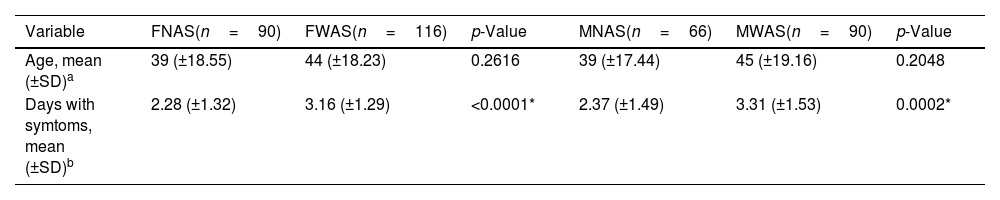
Participants were stratified into four groups based on sex and warning sign status: 90 women with dengue without warning signs (25%), 116 women with dengue with warning signs (32%), 66 men with dengue without warning signs (18%), and 90 men with dengue with warning signs (25%). This distribution revealed a higher proportion of female cases presenting with warning signs, potentially attributable to biological factors or differential exposure patterns (Table 1).

Table 1.

Gender-based differences in age and symptom duration among dengue patients with and without warning signs.

Variable  FNAS(n=90)  FWAS(n=116)  p-Value  MNAS(n=66)  MWAS(n=90)  p-Value 
Age, mean (±SD)a  39 (±18.55)  44 (±18.23)  0.2616  39 (±17.44)  45 (±19.16)  0.2048 
Days with symtoms, mean (±SD)b  2.28 (±1.32)  3.16 (±1.29)  <0.0001*  2.37 (±1.49)  3.31 (±1.53)  0.0002* 

Baseline characteristics of participants in the study. FNAS: female without warning signs; FWAS: female with warning signs; MNAS: male without warning signs; MWAS: male with warning signs. (a) Tukey's multiple comparisons test. (b) Student's t-test.

*

Significant differences p<0.05.

The mean age was 39 years for patients without warning signs and 44/45 years for those with warning signs. Comparative analysis using Student's t-test showed no significant between-group differences (p>0.05), suggesting age was not a determining factor for warning sign presentation in this cohort (Table 1).

Symptom duration (reported as time from symptom onset to hospital admission) showed clinically meaningful variations. Women without warning signs reported a mean duration of 2.28 days (±1.32), compared to 3.16 days (±1.29) for those with warning signs. Among male patients, these values were 2.37 days (±1.49) without warning signs versus 3.3 days (±1.53) with warning signs. These differences reached statistical significance (p<0.0001 for women; p=0.0002 for men). The extended pre-admission symptom duration observed in warning sign cases may reflect either slower disease progression or delayed healthcare-seeking behavior. These findings suggest the importance of maintaining close clinical surveillance, particularly when symptoms persist beyond the initial 48-h period (Table 1).

Hematological markers

Hematological parameters revealed significant alterations in patients with warning signs, demonstrating the dengue virus's impact on the hematopoietic system. Hematocrit levels showed a statistically significant increase in FWAS compared to FNAS at 24h (p<0.0001), 48h (p=0.0065), and 96h (p=0.0154) (Table 2). Similarly, MWAS exhibited elevated hematocrit at all timepoints compared to MNAS (24h: p<0.0001; 48h: p<0.0001; 72h: p=0.0002; 96h: p<0.0001). Moreover, men with warning signs had elevated hematocrit than women with warning signs at 24h (46.9±6.98% vs 44.5±4.83%, p<0.0335), 72h (44.2±7% vs 41.4±4.61%, p=0.0056), and 96h (43.4±6.99% vs 40.9±4.43%, p=0.0167), while no significant variations were observed between sexes in non-warning cases (Table 2).

Table 2.

Variations according to gender and time in hematological markers between patients with dengue with and without warning signs.

Hematological markers  Time(h)  FNAS(n=90)  FWAS(n=116)  MNAS(n=66)  MWAS(n=90)  p-Value
            FNAS vs FWAS  MNAS vs MWAS  FNAS vs MNAS  FWAS vs MWAS 
Hematocrit (%)Mean (±SD)24  41.3 (±5.22)  44.5 (±4.83)  41.6 (±5.84)  46. 9 (±6.98)  <0.0001*  <0.0001*  0.9843  0.0335* 
48  40.5 (±5.15)  42.9 (±5.01)  40.1 (±2.36)  44.8 (±6.87)  0.0065*  <0.0001*  0.8818  0.1335 
72  39.7 (±5.4)  41.4 (±4.61)  40.6 (±3.29)  44.2 (±7)  0.1123  0.0002*  0.62  0.0056* 
96  38.7 (±5.51)  40.9 (±4.43)  39.5 (±1.56)  43.4 (±6.99)  0.0154*  <0.0001*  0.5394  0.0167* 
Erythrocytes(×106/μL)Mean (±SD)24  4.7 (±0.51)  5 (±0.61)  5 (±0.64)  5.3 (±0.72)  0.0034*  0.0313*  0.1206  0.0539 
48  4.5 (±0.48)  4.8 (±0.44)  4.8 (±0.58)  5 (±0.48)  0.0002*  0.0303*  0.0107*  0.0011* 
72  4.4 (±0.43)  4.7 (±0.43)  4.7 (±0.69)  4.9 (±0.5)  <0.0001*  0.0386*  0.0491*  0.0007* 
96  4.4 (±0.41)  4.6 (±0.42)  4.7 (±0.52)  4.9 (±0.5)  0.0047*  0.0785  0.0024*  0.0002* 
Hemoglobin (g/dL)Mean (±SD)24  13.9 (±1.36)  14.6 (±1.65)  14.5 (±1.84)  15.7 (±1.95)  0.0094*  0.0004*  0.1669  <0.0001* 
48  13.6 (±1.38)  14.1 (±1.28)  14.1 (±1.7)  15.2 (±0.95)  0.0181*  <0.0001*  0.1844  <0.0001* 
72  13.4 (±1.31)  13.6 (±1.29)  14.3 (±1.92)  15 (±1.49)  0.3033  0.0887  0.0029*  <0.0001* 
96  13.4 (±1.34)  13.4 (±1.24)  13.8 (±1.63)  14.9 (±1.58)  0.9864  0.0003*  0.4807  <0.0001* 
Leukocytes(×103/μL)Mean (±SD)24  6.6 (±3.14)  4.6 (±2.57)  6.8 (±3.9)  4.8 (±2.39)  <0.0001*  0.0017*  0.9814  0.9286 
48  6.5 (±3.04)  4.9 (±2.53)  6.6 (±0.44)  4.8 (±3.14)  0.0006*  <0.0001*  0.9938  0.9956 
72  6.2 (±2.94)  5.3 (±2.52)  6.4 (±0.51)  5.2 (±1.89)  0.1043  <0.0001*  0.91  0.994 
96  5.9 (±2.91)  5.8 (±2.49)  6 (±0.41)  5.5 (±3.83)  0.9914  0.6007  0.9647  0.9514 
Lymphocytes(×103/μL)Mean (±SD)24  1.2 (±1.04)  1.2 (±0.9)  1.4 (±1.03)  1.3 (±0.93)  0.9994  0.9127  0.7533  0.9321 
48  1.5 (±0.96)  1.6 (±0.8)  1.6 (±0.64)  1.7 (±1)  0.8538  0.6055  0.9591  0.7633 
72  1.8 (±0.93)  1.9 (±0.72)  1.9 (±0.86)  2.2 (±1.32)  0.4818  0.3598  0.6777  0.2753 
96  2.1 (±0.86)  2.1 (±0.72)  2.2 (±1.09)  2.3 (±1.75)  0.9927  0.973  0.8382  0.7304 
Neutrophils(×103/μL)Mean (±SD)24  4.5 (±3.11)  2.4 (±1.58)  4.7 (±3.77)  2.4 (±1.88)  <0.0001*  <0.0001*  0.9854  >0.9999 
48  4.3 (±2.91)  1.9 (±1.52)  4.2 (±0.99)  1.9 (±1.63)  <0.0001*  <0.0001*  0.9862  0.998 
72  4.1 (±2.67)  1.9 (±1.37)  3.6 (±0.91)  1.9 (±1.88)  <0.0001*  <0.0001*  0.4294  0.9966 
96  3.6 (±2.49)  2.1 (±1.18)  2.7 (±0.73)  2.1 (±1.75)  <0.0001*  0.0079*  0.0113*  0.998 
Platelets(×103/μL)Mean (±SD)24  214 (±72.5)  61 (±55.1)  205 (±70.2)  44 (±32.3)  <0.0001*  <0.0001*  0.8479  0.0387* 
48  159 (±67.4)  43 (±38.7)  127 (±16.1)  32 (±16.2)  <0.0001*  <0.0001*  0.0003*  0.0352* 
72  129 (±56.2)  53 (±47.1)  111 (±16.7)  38 (±26.2)  <0.0001*  <0.0001*  0.0246*  0.0201* 
96  160 (±59.2)  69 (±60.3)  131 (±18.2)  49 (±30.5)  <0.0001*  <0.0001*  0.0001*  0.0136* 

Hematological values of study participants, stratified by time, sex and presence of warning signs. FNAS: female without warning signs; FWAS: female with warning signs; MNAS: male without warning signs; MWAS: male with warning signs. Tukey's multiple comparisons test: *Significant differences p<0.05.

Erythrocyte counts followed similar patterns, with significant increases in FWAS with respect to FNAS at 24h (p=0.0034), 48h (p=0.0002), 72h (p<0.0001) and 96h (p=0.0047), and in MWAS with respect to MNAS at 24h (p=0.0313), 48h (p=0.0303) and 72h (p=0.0386). Sex-based differences were evident, with men without warning signs showing higher counts than women without warning signs at 48h (4.8±0.44 vs 4.5±0.48×10⁶/μL, p=0.0107), 72h (4.7±0.69 vs 4.4±0.43×10⁶/μL, p=0.0491), and 96h (4.7±0.52 vs 4.4±0.41×10⁶/μL, p=0.0024) (Table 2). Hemoglobin concentrations mirrored these findings, with significant elevations in warning cases and consistent sex differences.

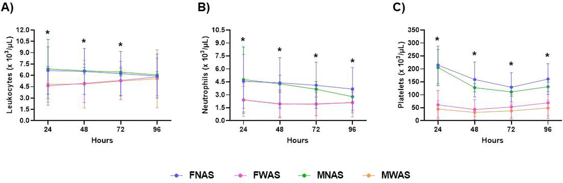
Leukocyte counts demonstrated significant decreases in cases with warning signs respect to without warning signs cases. In this sense, women showing leukopenia at 24h (4.6±2.57×10³/μL, p<0.0001) and 48h (4.9±2.53×10³/μL, p=0.0006), and men at 24h (4.8±2.39×10³/μL, p=0.0017), 48h (4.8±3.14×10³/μL, p<0.0001), and 72h (5.2±3.83×10³/μL, p<0.0001) (Table 2) (Fig. 1A). Neutropenia was particularly pronounced, affecting both sexes with warning signs at all timepoints (p<0.0001) (Table 2 and Fig. 1B). In contrast, lymphocyte counts remained stable across all groups and timepoints (p>0.05) (Table 2).

Fig. 1.

Longitudinal changes in hematological markers by dengue severity and sex. Time-course analysis of (A) leukocytes, (B) neutrophils, and (C) platelets in dengue patients, stratified by sex and clinical severity. Data represent mean±standard deviation. Groups: FNAS (females without warning signs), FWAS (females with warning signs), MNAS (males without warning signs), and MWAS (males with warning signs). Significant differences between groups at specific time points (Tukey's post-hoc test, p<0.05) are indicated with asterisks (*).

Thrombocytopenia showed progressive development, with significant platelet reductions in FWAS compared with FNAS at 24h (61±55.1×10³/μL vs 214±72.5×10³/μL, p<0.0001), 48h (43±38.7×10³/μL vs 159±67.4×10³/μL, p<0.0001), 72h (53±47.1×10³/μL vs 129±56.2×10³/μL, p<0.0001), and 96h (69±60.3×10³/μL vs 160±59.2×10³/μL, p<0.0001), and similar patterns in men (Table 2). Sex differences were notable, with men consistently showing lower platelet counts than women in both warning and non-warning groups at most timepoints (Fig. 1C).

The hematological findings demonstrate significant alterations in dengue patients with warning signs, characterized by marked hemoconcentration (elevated hematocrit, erythrocytes, and hemoglobin), progressive thrombocytopenia, and leukopenia with pronounced neutropenia. These patterns, which exhibit sex-specific variations – particularly more severe hematocrit elevation and platelet reduction in males – suggest plasma leakage and possible bone marrow suppression during the critical phase. The stability of lymphocyte counts contrasts with other cell line disturbances, implying selective hematopoietic involvement. These results highlight the potential clinical utility of serial hematocrit and platelet monitoring, particularly in male patients, as part of early risk stratification. The observed sex disparities warrant further investigation into possible immunological or hormonal influences on dengue progression.

Hepatic function markers

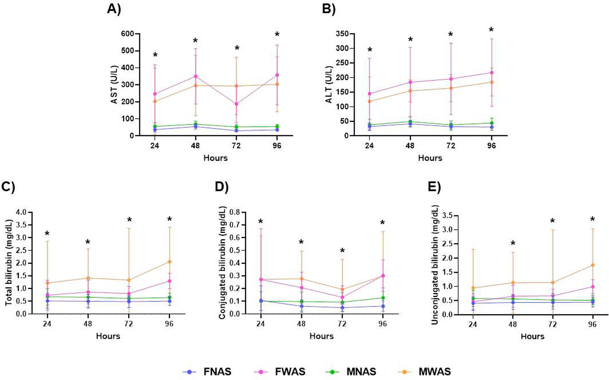
The analysis revealed significant AST elevations in women with warning signs (24h: 247.2±170.4U/L, p<0.0001; 48h: 350±162.6U/L, p<0.0001; 72h: 187.6±109.7U/L, p<0.0001; 96h: 358.1±175.5U/L, p<0.0001) compared to women with non-warning sings. Men showed parallel increases (24h: 203.1±195.1U/L, p<0.0001; 48h: 296.4±177.5U/L, p<0.0001; 72h: 293±169.1U/L, p<0.0001; 96h: 303.7±161.4U/L, p<0.0001) compared to men with non-warning sings (Table 3), with baseline sex differences evident (men without warning signs vs women without warning signs at all timepoints, p<0.0001). The most notable sexual dimorphism occurred at 72h, when men with warning signs (293±169.1U/L) surpassed women with warning signs (187.6±109.7U/L, p<0.0001) (Table 3).

Table 3.

Hepatic markers in dengue: differential patterns by gender, time and clinical severity.

Liver function markers  Time(h)  FNAS(n=90)  FWAS(n=116)  MNAS(n=66)  MWAS(n=90)  p-Value
            FNAS vs FWAS  MNAS vs MWAS  FNAS vs MNAS  FWAS vs MWAS 
AST (U/L)Mean (±SD)24  36.9 (±13.96)  247.2 (±170.4)  55.7 (±22.6)  203.1 (±195.1)  <0.0001*  <0.0001*  <0.0001*  0.3252 
48  54.8 (±15.84)  350 (±162.6)  69.4 (±17.17)  296.4 (±177.5)  <0.0001*  <0.0001*  <0.0001*  0.1190 
72  30.8 (±7.75)  187.6 (±109.7)  52.5 (±13.16)  293 (±169.1)  <0.0001*  <0.0001*  <0.0001*  <0.0001* 
96  35 (±7.37)  358.1 (±175.5)  54.9 (±13.35)  303.7 (±161.4)  <0.0001*  <0.0001*  <0.0001*  0.0999 
ALT (U/L)Mean (±SD)24  32.1 (±12.72)  144.7 (±121.5)  37.9 (±19.55)  118.5 (±83.39)  <0.0001*  <0.0001*  0.1553  0.2638 
48  41.7 (±11.23)  184.1 (±119.6)  49.2 (±16.65)  154.5 (±38.03)  <0.0001*  <0.0001*  0.0053*  0.0635 
72  31.5 (±12.55)  195.7 (±122.2)  37.8 (±13.91)  163.7 (±46.6)  <0.0001*  <0.0001*  0.0212*  0.0519 
96  30.2 (±10.73)  217.1 (±115.4)  44 (±16.64)  184.4 (±48.02)  <0.0001*  <0.0001*  <0.0001*  0.0324* 
Total bilirubin(mg/dL)Mean (±SD)24  0.51 (±0.293)  0.73 (±0.59)  0.67 (±0.324)  1.22 (±1.641)  0.0028*  0.0146*  0.0082*  0.0425* 
48  0.5 (±0.266)  0.86 (±0.466)  0.66 (±0.188)  1.41 (±1.143)  <0.0001*  <0.0001*  0.0001*  0.0002* 
72  0.48 (±0.233)  0.8 (±0.282)  0.61 (±0.276)  1.33 (±2.028)  <0.0001*  0.0068*  0.0144*  0.0739 
96  0.5 (±0.165)  1.29 (±0.304)  0.64 (±0.179)  2.05 (±1.35)  <0.0001*  <0.0001*  <0.0001*  <0.0001* 
Conjugated bilirubin(mg/dL)Mean (±SD)24  0.1 (±0.116)  0.27 (±0.399)  0.1 (±0.73)  0.27 (±0.342)  0.0002*  <0.0001*  0.9916  >0.9999 
48  0.06 (±0.049)  0.2 (±0.122)  0.09 (±0.075)  0.27 (±0.218)  <0.0001*  <0.0001*  0.0062*  0.0352* 
72  0.05 (±0.029)  0.13 (±0.09)  0.09 (±0.075)  0.19 (±±0.237)  <0.0001*  0.0015*  0.0002*  0.0921 
96  0.06 (±0.04)  0.3 (±0.124)  0.12 (±0.079)  0.3 (±0.348)  <0.0001*  <0.0001*  <0.0001*  >0.9999 
Unconjugated bilirubin(mg/dL)Mean (±SD)24  0.4 (±0.251)  0.46 (±0.278)  0.57 (±0.286)  0.94 (±1.369)  0.4079  0.0637  0.0012*  0.0073* 
48  0.43 (±0.254)  0.65 (±0.395)  0.56 (±0.163)  1.13 (±1.082)  <0.0001*  <0.0001*  0.0017*  0.0007* 
72  0.43 (±0.231)  0.67 (±0.229)  0.52 (±0.233)  1.14 (±1.862)  <0.0001*  0.0125*  0.1102  0.0912 
96  0.44 (±0.172)  0.99 (±0.249)  0.52 (±0.164)  1.75 (±1.283)  <0.0001*  <0.0001*  0.0367*  <0.0001* 

Liver function markers in study participants, categorized by time, sex and disease severity. FNAS: female without warning signs; FWAS: female with warning signs; MNAS: male without warning signs; MWAS: male with warning signs. Tukey's multiple comparisons test: *Significant differences p<0.05.

ALT profiles showed similar patterns to AST, with warning cases showing progressive elevations (women, 24h: 144.7±121.5U/L, p<0.0001 to 96h: 217.1±115.4U/L, p<0.0001; men, 24h: 118.5±83.39U/L, p<0.0001 to 96h: 184.4±48.02U/L, p<0.0001). Sex-based divergence peaked at 96h (women with warning signs: 217.1±115.4U/L vs men with warning signs: 184.4±48.02U/L, p=0.324), while in cases without alarm symptoms, a lower concentration of aspartate aminotransferase was observed in women repect to men at 48h (p=0.0053), 72h (p=0.0212) and 96h (p<0.0001) (Table 3).

Total bilirubin showed sustained increases in warning cases (women: [24h: 0.73±0.59mg/dL, p=0.0028] to [96h: 1.29±0.304mg/dL, p<0.0001]; men: [24h: 1.22±1.641mg/dL, p=0.0146] to [96h: 2.05±1.35mg/dL, p<0.0001]). In this sense, both conjugated bilirubin and unconjugated bilirubin, showed significant increases in patients with warning signs compared to patients without warning signs (Table 3). A significant increase was also observed in men compared to women for both types of bilirubin.

The trajectories of liver parameters show three clinically relevant patterns: first, a consistent progression of liver injury markers over the clinical course, rather than isolated peaks; second, sexually dimorphic responses suggesting that male patients may have greater baseline liver vulnerability, progressing to more progressive damage after the onset of warning signs; and third, distinct temporal patterns among bilirubin fractions, indicating phase-specific pathophysiological mechanisms. These findings, taken together, suggest that liver monitoring protocols should incorporate1: sex-specific reference ranges for interpretation2; differential alert thresholds for conjugated and unconjugated bilirubin fractions; and3 expanded surveillance windows to detect late-onset dysfunction, particularly in women who present with late but significant enzyme elevations. Bilirubin metabolism patterns further suggest the need to assess biliary excretory function along with hepatocellular injury in severe cases of dengue (Fig. 2).

Fig. 2.

Longitudinal changes in liver function markers by dengue severity and sex. Time-course analysis of liver function markers in dengue patients, stratified by sex and clinical severity: (A) AST, (B) ALT, (C) total bilirubin, (D) conjugated bilirubin, and (E) unconjugated bilirubin. Data represent mean±standard deviation. Groups: FNAS (females without warning signs), FWAS (females with warning signs), MNAS (males without warning signs), and MWAS (males with warning signs). Significant differences between groups at specific time points (Tukey's post-hoc test, p<0.05) are indicated with asterisks (*).

Renal function markers

Serum urea concentrations showed no statistically significant differences between women with and without warning signs at any evaluated timepoint (p>0.05). Similarly, no significant variations were observed in men with warning signs compared to those without across all timepoints (p>0.05). However, sex-based differences were evident: men without warning signs exhibited higher urea levels than women without warning signs at 24h (p<0.0001), 48h (p<0.0001), 72h (p=0008) and 96h (p=0.0015). This pattern persisted in warning cases, with men showing elevated urea compared to women at 24h (p=0.0058), 48h (p=0.0075), 72h (p=0096) and 96h (p=0.0078) (Table 4).

Table 4.

Time and sex-specific kidney function profiles in dengue.

Kidney function markers  Time(h)  FNAS(n=90)  FWAS(n=116)  MNAS(n=66)  MWAS(n=90)  p-Value
            FNAS vs FWAS  MNAS vs MWAS  FNAS vs MNAS  FWAS vs MWAS 
Urea (mg/dL)Mean (±SD)24  23.2 (±14.08)  26.9 (±15.52)  34.6 (±16.02)  40.1 (±34.88)  0.2772  0.5487  <0.0001*  0.0058* 
48  22.1 (±13.4)  23.9 (±10.39)  32.4 (±9.32)  35 (±30.71)  0.7116  0.8806  <0.0001*  0.0075* 
72  19.3 (±11.74)  20.7 (±8.172)  25.4 (±7.57)  30.7 (±28.76)  0.7783  0.3331  0.0008*  0.0096* 
96  18.6 (±11.33)  19.8 (±6.918)  24.68 (±8.72)  29.7 (±28.11)  0.8370  0.3767  0.0015*  0.0078* 
Creatinine (mg/dL)Mean (±SD)24  0.7 (±0.355)  0.75 (±0.754)  1.45 (±1.178)  1.26 (±1.265)  0.9189  0.7739  <0.0001*  0.0056* 
48  0.94 (±0.393)  0.69 (±0.329)  1.67 (±1.369)  1.08 (±1.165)  <0.0001*  0.0292*  0.0004*  0.0114* 
72  0.61 (±0.253)  0.72 (±0.237)  0.84 (±0.763)  0.89 (±1.155)  0.0077*  0.9849  0.0992  0.5188 
96  0.66 (±0.233)  0.71 (±0.244)  0.83 (±0.85)  0.85 (±1.057)  0.5404  0.9986  0.4239  0.586 

Kidney function markers in study participants, classified by time, sex and clinical status. FNAS: female without warning signs; FWAS: female with warning signs; MNAS: male without warning signs; MWAS: male with warning signs. Tukey's multiple comparisons test: *Significant differences p<0.05.

Creatinine dynamics revealed more complex alterations. FWAS displayed a significant decrease compared to FNAS at 48h (0.69±0.329mg/dL vs 0.94±0.393mg/dL, p<0.0001) but an increase at 72h (0.72±0.237mg/dL vs 0.61±0.253mg/dL, p=0.0077). MWAS similarly showed reduced creatinine at 48h (1.08±1.165mg/dL vs 1.67±1.369mg/dL, p=0.0292) compared with MNAS. Sex disparities were prominent: MNAS had higher creatinine than FNAS at 24h (p<0.0001) and 48h (p=0.0004), a trend mirrored in warning groups, 24h (p=0.0056) and 48h (p=0.0114) (Table 4).

While these findings suggest preserved baseline renal function in most patients, the transient creatinine fluctuations – particularly the 48h decrease in both sexes with warning signs followed by a 72h rebound in women – may reflect early hemodynamic or metabolic shifts warranting further study. The consistent sex differences in urea and creatinine, independent of warning signs, highlight potential physiological or hormonal influences on renal biomarkers during dengue infection.

Discussion

Our study findings, consistent with recent research by Ahmad et al. in Southeast Asia and the retrospective study of Hernández-Bautista et al., support the need for differentiated monitoring protocols for dengue patients presenting warning signs.16,17 Unlike previous studies that primarily focused on platelet counts, our results – mirroring those of Sangkaew et al. – support the importance of evaluating multiple parameters at specific timepoints for more accurate risk stratification,18 as the clinical evolution of patients diagnosed with severe dengue is closely related to changes in hematological biomarkers and those related to liver function. Monitoring these trends is essential for proper risk stratification and real-time clinical decision-making. Reported studies highlight the importance of monitoring certain hematological and hepatic markers during the critical phases of the disease with the aim of early identification of patients at risk of severe complications.

In patients with dengue, the persistence of warning signs beyond 48h is associated with a higher risk of hematological and hepatic deterioration and should be a critical stratification point for clinical management. During the initial 48-h febrile phase, we recommend an approach similar to IDSA guidelines but modified based on our findings.19 While WHO traditionally emphasizes platelet and hematocrit monitoring – the most commonly used parameters to assess disease severity – our data demonstrate that combining these with neutrophil counts and early liver enzymes (AST/ALT) may improve detection of patients likely to progress to severe disease.9 A progressive decline in platelets below 100,000/μL and an increase in hematocrit indicate a change in capillary permeability, a phenomenon characteristic of severe dengue and related to capillary leak syndrome; in the context of persistent warning signs, this should be considered an indicator of high risk for dengue shock syndrome (DSS) and hemorrhage. This partially aligns with findings from Nguyen et al. in Vietnam, though our study revealed more pronounced liver involvement patterns in female patients – an aspect not previously reported.20

The 48–72-h critical window requires particular attention, as noted by WHO dengue guidelines.9 However, unlike studies recommending 24-h monitoring intervals, our results suggest that patients with persistent warning signs or worsening initial markers may require 12-h assessments. This recommendation stems from our observation of rapid hematological changes, which may signal imminent clinical deterioration. Hepatic markers, such as AST/ALT, have been identified as key indicators of liver damage in this pathology. This study confirms that a progressive increase in AST and ALT levels, especially when AST exceeds 500U/L – a sign of liver deterioration that may precede liver failure – is directly related to a higher risk of progression to severe dengue.

For the subsequent monitoring period (72–168h), we propose incorporating bilirubin fractionation based on our findings on sex-specific patterns of liver involvement. The analysis of gender differences in the evolution of biomarkers is also relevant for clinical decision-making. This study shows that women with warning signs exhibit more pronounced patterns of liver deterioration, which strengthens the need to implement sex-differentiated monitoring protocols in patients with severe dengue. In this regard, our findings suggest that certain subgroups (particularly patients with comorbidities or receiving intensive fluid therapy) could benefit from specific assessments of renal function during this phase.

Implementing this comprehensive monitoring approach, which combines established guidelines with our new findings, could address the limitations of current protocols. As proposed by Raafat et al., they suggest implementing monitoring strategies that incorporate multiple parameters and demographic differences to achieve better early identification of cases likely to progress to severe disease.21 The combination of hematological and hepatic markers can serve as crucial reference points for critical clinical decisions, such as intensifying monitoring or hospitalizing patients. In clinical settings, the results of these biomarkers are essential for determining management; for example, monitoring platelet count and hematocrit may guide adjustments in intravenous therapy, while an upward trend in AST/ALT levels may justify referral to intensive care units.

Our findings provide a clear evidence-based rationale for using the 48-h symptom mark as a critical triage tool in endemic settings. Patients whose warning signs persist beyond this point are at significantly higher risk of complications and should be considered for extended observation, hospital admission, or more intensive monitoring protocols. Conversely, the absence of warning signs after 48h, coupled with stable laboratory trends, could support safer early discharge decisions, optimizing resource allocation in overburdened healthcare systems. These evidence-based recommendations represent a balanced integration of current information and clinical feasibility, with the potential to reduce severe dengue complications through early risk identification. The integration of these clinical indicators and biomarkers not only helps improve clinical outcomes but also optimizes the use of resources in hospitals and primary care units. Future research should validate these protocols in diverse epidemiological contexts and assess their impact on clinical outcomes, paying special attention to the implementation of sex-specific monitoring approaches.

Ethics declarations

This study was conducted in accordance with the Declaration of Helsinki (1976). It was also revised and approved by the Research and Ethics Committees of the Instituto Mexicano de Seguridad Social, with the registration number 28.202. Informed consent was obtained from all participants. All patients included in this study were informed in writing regarding the collection of their samples for research aims and were given the right to refuse participation.

Conflict of interest

The authors declare that they have no known competing financial interests or personal relationships that could have appeared to influence the work reported in this paper. No funding sources or entities with potential conflicts were involved in the design, execution, analysis, or interpretation of this study.

Appendix B
Supplementary data

The following are the supplementary data to this article:

Icono mmc1.doc

References
[1]
M.T. de Almeida, D.G. Merighi, A.B. Visnardi, et al.
Latin America's dengue outbreak poses a global health threat.
Viruses, 17 (2025),
[2]
S.J. Ryan, C.J. Carlson, E.A. Mordecai, L.R. Johnson.
Global expansion and redistribution of Aedes-borne virus transmission risk with climate change.
PLoS Negl Trop Dis, 13 (2019),
[3]
World Healt Organization.
Dengue and Severe Dengue [Internet].
WHO, (2025),
[4]
Pan American Health Organization.
Dengue Outbreaks in Latin America: Health and Economic Impacts [Internet].
PAHO, (2025),
[5]
Secretaría de Salud.
Panorama epidemiológico de fiebre por dengue y fiebre hemorrágica por dengue con información del Sistema Especial de Vigilancia Epidemiológica de Dengue: Publicación por semana epidemiológica a cargo de la Dirección de Vigilancia Epidemiológica de Enfermedades Transmisibles (Semana epidemiológica 52 del, 2024) [Internet].
Dirección de Vigilancia Epidemiológica de Enfermedades Transmisibles, (2024),
[6]
P. Sankoju, V.S.U. Ravinuthala, R. Mopuri, S.R. Mutheneni, A. Addlagatta.
Genomic characterization and evolutionary analysis of dengue virus from Aedes mosquitoes in Telangana, India.
J Vector Borne Dis, 60 (2023), pp. 179-186
[7]
P.F. Hernández Bautista, D.A. Cabrera Gaytán, C.E. Santacruz Tinoco, et al.
Retrospective analysis of severe dengue by dengue virus serotypes in a population with social security, Mexico 2023.
Viruses, 16 (2024),
[8]
M. Riaz, S.N.B. Harun, T.H. Mallhi, et al.
Evaluation of clinical and laboratory characteristics of dengue viral infection and risk factors of dengue hemorrhagic fever: A multi-center retrospective analysis.
BMC Infect Dis, 24 (2024), pp. 500
[9]
World Healt Organization.
Dengue: Guidelines for Diagnosis, Treatment, Prevention and Control [Internet].
WHO, (2009),
[10]
F. Narvaez, G. Gutierrez, M.A. Pérez, et al.
Evaluation of the traditional and revised WHO classifications of dengue disease severity.
PLoS Negl Trop Dis, 5 (2011), pp. e1397
[11]
World Health Organization, UNICEF. Handbook for Clinical Management of Dengue; 2012.
[12]
A.M. Swamy, P.Y. Mahesh, S.T. Rajashekar.
Liver function in dengue and its correlation with disease severity: a retrospective cross-sectional observational study in a tertiary care center in Coastal India.
Pan Afr Med J, 40 (2021),
[13]
P. Gurugama, U. Jayarajah, K. Wanigasuriya, A. Wijewickrama, J. Perera, S.L. Seneviratne.
Renal manifestations of dengue virus infections.
J Clin Virol, 101 (2018), pp. 1-6
[14]
J.K. Paul, M. Azmal, T. Alam, O.F. Talukder, A. Ghosh.
Comprehensive analysis of intervention and control studies for the computational identification of dengue biomarker genes.
PLoS Negl Trop Dis, 19 (2025),
[15]
T.J. Schaefer, P.K. Panda, R.W. Wolford.
Dengue fever.
StatPearls [Internet], StatPearls Publishing, (2024),
[16]
L.C.R.Q. Ahmad, B.S. Gill, L.H. Sulaiman, et al.
Molecular epidemiology of dengue in Southeast Asia (SEA): protocol of systematic review and meta-analysis.
BMJ Open, 15 (2025),
[17]
P.F. Hernández Bautista, D.A. Cabrera Gaytán, C.E. Santacruz Tinoco, et al.
Retrospective analysis of severe dengue by dengue virus serotypes in a population with social security, Mexico 2023.
Viruses, 16 (2024), pp. 769
[18]
S. Sangkaew, D. Ming, A. Boonyasiri, et al.
Risk predictors of progression to severe disease during the febrile phase of dengue: A systematic review and meta-analysis.
Lancet Infect Dis, 21 (2021), pp. 1014-1026
[19]
J.M. Miller, M.J. Binnicker, S. Campbell, et al.
IDSA Guidelines.
(2024),
[20]
R.N. Nguyen, H.T. Lam, H.V. Phan, H.T. LAM, H.V. PHAN.
Liver impairment and elevated aminotransferase levels predict severe dengue in Vietnamese children.
Cureus, 15 (2023),
[21]
N. Raafat, S. Loganathan, M. Mukaka, S.D. Blacksell, R.J. Maude.
Diagnostic accuracy of the WHO clinical definitions for dengue and implications for surveillance: A systematic review and meta-analysis.
PLoS Negl Trop Dis, 15 (2021),
Copyright © 2025. Sociedad Española de Enfermedades Infecciosas y Microbiología Clínica
asdasdasd
Opciones de artículo
Herramientas
Material suplementario