
Herlyn–Werner–Wünderlich syndrome is a uterine malformation characterized by uterus didelphys, obstructed hemivagina and ipsilateral renal agenesis.
Clinical findingsThe manifestation of the disease is widely diverse; it is usually diagnosed after menarche, with dysmenorrhea and abnormal uterine bleeding; it is also associated with infertility.
Main diagnosisFour clinical cases, their diagnosis are reported here.
Therapeutic interventions and resultsThe treatment and results of these four patients are described here.
ConclusionWhen studying uterine malformation it is important to consider this rare disease to avoid possible complications and giving the patient a correct diagnose and treatment. The hysteroscopy resection of the longitudinal vaginal septum in those symptomatic patients with hematocolpos should be considered as a good option for treatment.
El síndrome de Herlyn-Werner-Wünderlich es una malformación uterina que asocia útero didelfo, hemivagina obstruida total o parcialmente y agenesia renal ipsilateral.
Hallazgos clínicosLa clínica que presenta este síndrome es muy diversa; se suele diagnosticar después de la menarquia cursando con dismenorrea y sangrado uterino anómalo; así mismo se asocia a infertilidad.
Diagnósticos principalesSe presentan a continuación 4 casos clínicos, su diagnóstico y tratamiento mediante diversas técnicas.
Intervenciones terapéuticas y resultadosSe describen en este manuscrito los tratamientos aplicados a estas pacientes y sus resultados.
ConclusiónAnte el hallazgo de una malformación uterina es importante tener en cuenta esta entidad infrecuente, para evitar posibles complicaciones y proporcionar a la paciente un diagnóstico y tratamiento correctos. La resección histeroscópica del tabique vaginal longitudinal en aquellas pacientes sintomáticas con hematocolpos debe ser considerada como una buena opción de tratamiento.

The double uterus malformation or uterus didelphys is rarely associated with unilateral obstructed hemivagina and ipsilateral renal agenesis.1 The term “Herlyn–Werner–Wunderlich” was mentioned in 19792 to define the triad anomaly in a report of four urogenital anomalies cases, they had in common the presence of a double uterus, a paracervical cyst and ipsilateral renal aplasia. The current acronym is OHVIRA (Obstructed Hemivagina and Ipsilateral Renal Anomaly), proposed by Smith et al., in 2007.3-5
During the embryonic, the development of Müllerian structures (uterus, vagina and Fallopian tubes) is independent of the gonadal structures; on the other hand, it is related with the urinary system; that is the reason why it is usual for Mülllerian anomalies to associate urinary or renal anomalies and not gonadal anomalies. This anomaly is due to a defect of the ducts of Müller.
It is often diagnosed at menarche, usually with dysmenorrhea, cyclic pelvic pain as from a hematocolpos/hematometrocolpos, originated by a septate vagina. Ultrasound and MR (magnetic resonance) are especially useful to identify the anomalies. A correct identification of the syndrome enables the treatment, usually being surgical excision of the vaginal septum. This surgery provides a relief from the symptoms and prevention of additional complications derived from the syndrome. Since multicystic dysplasia and renal agenesis are commonly diagnosed on prenatal or neonatal period, most authors recommend checking the possibility of associated Müllerian duct anomalies.6,7
The most widely used classification systems are that of the American Fertility Society, (currently the American Society for Reproductive Medicine) created in 1988 and revised in 2021, which establishes nine types; and the classification of the European Society of Human Reproduction and Embryology and the European Society of Gastrointestinal Endoscopy of 2013 (Table 1, supplementary data). The main difference between them is while the first classifies only the uterine body, the second classifies body, cervix and vagina independently; so a complete diagnosis can be made by simplifying the nomenclature.
Four cases of this syndrome have been described in which it has been taken in consideration its clinical, diagnostic and therapeutic features. The protocols of the University Hospital on the publication of patient data have been followed, patient privacy has been respected, and we have the consent of the patients for publication.
Patient informationCase 1: A 26-years-old woman came to our emergency Department for persistent left iliac fossa pain, which irradiated to the bottom of the abdomen. Apparently, it was a healthy woman, any clinical background. She had dysmenorrhea and she had never been pregnant.
Case 2: A 16-years-old woman, redirected from Internal Medicine for a gynecologic identification owing to suspecting a uterine malformation.
Case 3: A 21-years-old patient came with dysmenorrhea, stink flux and intermenstrual bleeding. About her personal background, she had a renal agenesis diagnosed in childhood and a current diagnostic of double uterus and double cervices.
Case 4: A 16-years-old woman, which case was followed up owing to a double uterus and cervices on childhood and right renal agenesia. She did not show any associated symptoms.
Clinical findingsCase 1: The patient was diagnosed with uterus didelphys after a hysterosalpingography test realized in a basic fertility exam some years ago, which showed an uterus with an elongated morphology, smooth walls, right-turned, and since the contrast reached the peritoneal cavity, the fallopian tube was permeable.
Case 2: The patient arrived with symptoms matching pyelonephritis, thus a computed abdomen–pelvic tomography was performed, which showed uterine malformation. Aside from the symptoms of pyelonephritis, only dysmenorrhea in a low-moderate level was found as symptoms with a gineco-obstetric origin.
Case 3: The patient had normal female external genitalia were showed but a protuberance in the left side of the vagina which matched with hematocolpos in the septate vagina. A single and normal cervix was observed. Transvaginal ultrasound showed a double uterus (both were normal), which and left endometrial thickness of 7mm and right of 8mm. A single cervix and cyst, which might belong to a hematocolpos in a septate vagina, were visualized. In the fourth case a hysteroscopy was performed at 13 years old (with a draining of the septate vagina) and a cystoscopy owing to a ureteral reflux.
Case 4: a hysteroscopy was performed at 13 years old (with a draining of the septate vagina) and a cystoscopy owing to a ureteral reflux. External genitalia were normal, no speculoscopy for whole hymen was performed.
TimelineYounger patients (cases 2 and 4) remained asymptomatic, but patients in cases 1 and 3 had dysmenorrhea, infertility, and stink flux.
Diagnostic evaluationCase 1: A computed abdomen–pelvic tomography was made. It revealed left renal agenesis and right renal hypertrophy of 13cm without any injury or dilatation associated.
In a gynecologic exploration, the external genitalia and the vagina was fine aside from some slightly stinky menstruation remnants. A single external cervical orifice was observed, well epithelized.
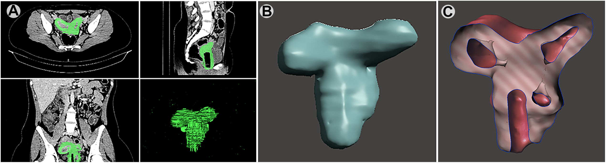
The abdomen–pelvic TC showed bicornuate uterus, two uterine bodies and two cervices separated by a longitudinal vaginal septum. It was observed a 12mm cyst in the left hemivagina. There was neither hematocolpos nor hematometra (Fig. 1A). Thus, the patient was diagnosed with OHVIRA syndrome due to the addition of renal agenesis and septate hemivagina to her already know uterine malformation.
(A) CT scan of the pelvis, selection of the structures. (B) 3D virtual reconstruction of the selected structures (double uterus and vaginas). (C) Middle section of the uterus showing double uterus, and one opened vagina and the obliterated vagina. (D) Resection of the longitudinal vaginal septum by vaginoscopic.
In this case, a conversion procedure was applied to transform the CT (computed tomography) scan into a 3D model.8 CT scans in a digital imaging and communications in medicine (DICOM) format were imported to the imaging software InVesalius v3.18 and rendered to generate virtual reconstructions of the selected structures (Fig. 1B).
The use of InVesalius generated 3D surfaces in standard triangulation language (STL). The meshes generated were corrected using MeshLab v2016.12,9 open-source software that enables cleaning, remodeling, and remeshing of the structure. The use of MeshLab is essential to make the 3D surface hollow, and consequently, lighter. The structure obtained with MeshLab have holes, cavities, and, in general, imperfections. In order to remove them, another free 3D modeling software called Meshmixer v2017 was used10 (Fig. 1C).
Case 2: For the gynecologic examination, since speculoscopy was discarded due to the integrity of the hymen, an abdominal ultrasound was performed. It revealed an anteverted bicornuate uterus with a single cervix. The endometrial thickness was 14mm right and 10mm left. Both ovaries had a regular look. Right unique kidney with 3 stones, adjacent to the double J catheter was visualized.
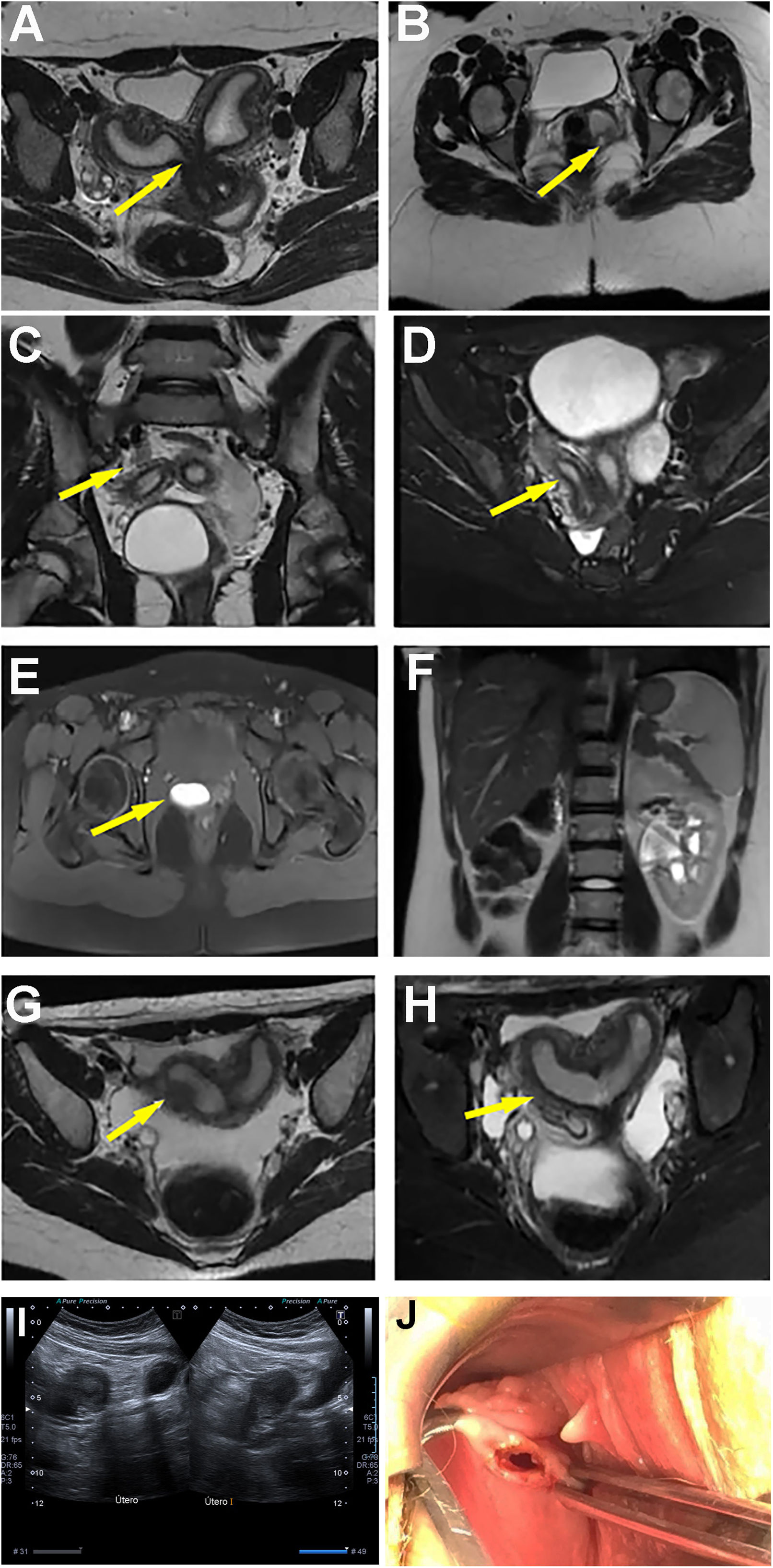
Pelvic MR was performed to complete the study whose report described left renal agenesis, two cervices separated, one rudimentary cervix that ends in a blind vagina and the other cervix continues into a bicornuate uterus (Fig. 2A, B).
(A) Pelvic MR, sequential axial cuts T2: it is identified a uterus didelphys with a longitudinal vaginal septum (arrow). (B) Presence of two hemivaginas. Enlarged left hemivagina and with hyperintense content as regards hematocolpos (arrow). (C) Uterus didelphys (arrow). (D) Arrow shows hyperintense content in the left hemivagina (hematocolpos). (E) Cystic image of the postero-lateral left wall of the bladder regarding the ureterocele (arrow). (F) Left renal agenesis. (G and H) pelvic MR, sequences T2 axial and coronal; uterus didelphys (arrow). (I) Abdominal ultrasound showing the double uterus. (J) Resection of the longitudinal vaginal septum by vaginoscopic.
This case, since there was no possible the proper exploration to find hematocolpos or hematometra, could be a Herlyn–Werner–Wünderlich syndrome rather than an OHVIRA.
Case 3: As previously, to complete the study, a pelvic MR was performed, which showed left renal agenesis, uterus didelphys and a longitudinal vaginal septum and left hematocolpos explaining the septate vagina (the left one seemed to be filled and isolated from the outside) (Fig. 2C–F). The patient was appropriate to be diagnosed as OVHIRA syndrome.
Case 4: The abdominal ultrasound showed a uterus on the left side measures of 69mm×36mm×27mm. However, it also showed a second uterus, of smaller size (60×34×30) (Fig. 2I). Taking into consideration this new observation and the renal agenesis background, it was restored a previous pelvic MR, in which was described right renal agenesis (with compensatory hypertrophy on the left side), uterus didelphys and two vaginas. On the right side of the vagina, liquid accumulation could be appreciated, with measures of 4.2mm×3.8mm×8.1mm (being compatible with hematic content) and the left vaginal cavity was collapsed and empty of liquid (Fig. 2G–H). Thus, the patient was diagnosed with OHVIRA syndrome with hematocolpos on the right septate vagina.
Therapeutic interventionCase 1: The treatment was a resection of the longitudinal vaginal septum by vaginoscopic access and exposition of the septate hemivagina, providing a proper evacuation in both cavities (Fig. 1D).
Case 2: No surgery has been necessary, but the dysmenorrhea has been treated.
Case 3: The treatment was a surgery by vaginoscopic use of a resectoscope to remove the longitudinal vaginal septum, then draining and communicate the septate hemivagina with the rest of the vagina and the outside.
Case 4: The patient undergone surgery by vaginoscopic hysteroscopy for vaginal septum resection and draining the hematocolpos (Fig. 2J), connecting both vagina and both cervices.
Follow up and resultsAll patients were followed up in consultation with regular clinical monitoring after diagnosis to assess the evolution.
Case 1: The patient got pregnant spontaneously, concluding as a planned cesarean section due a fetal gastroschisis.
Case 2: No surgery has been necessary, but the dysmenorrhea has been treated.
Case 3: On later medical appointments, the patient was asymptomatic and with a non-complicated pregnancy, which finished as a cesarean section.
Case 4: Has a good evolution.
DiscussionMost of publications focus on the reproductive capabilities of patients with the anomaly, but do not provide insight about gynecologic problems (such as endometriosis).6,7 OHVIRA syndrome, as far as we identify, is not associated to primary infertility, and the pregnancy can go without surgery. The evidence does not support nowadays the hysteroscopy resection of the longitudinal vaginal septum for asymptomatic patients or before the first pregnancy but in our experience seems to be a good option for patients with hematocolpos.
Ethical disclosuresProtection of people and animalsThe authors declare that no experiments have been carried out on humans or animals for this research.
Confidentiality of dataThe authors declare that they have followed their workplace's protocols regarding the publication of patient data.
Right to privacy and informed consentThe authors have obtained informed consent from the patients and/or subjects referred to in the article. This document is in the possession of the corresponding author.
Ethics and consent for publicationThe protocols of the University Hospital of Puerto Real (Cadiz, Spain) on the publication of patient data have been followed.
The patient privacy has been respected.
The researchers obtained consent for publication from the patients.
FundingThis research received no specific grant from any funding agency in the public or commercial sectors.
Conflict of interestThe authors declare that they have no conflict of interests.