metricas
covid
Cirugía Española (English Edition) Primary axillary hydatid cyst: A rare location
Journal Information
Vol. 99. Issue 6.
Pages 466 (June - July 2021)
Vol. 99. Issue 6.
Pages 466 (June - July 2021)
Image of the month
Full text access
Primary axillary hydatid cyst: A rare location
Quiste hidatídico axilar primario: una localización rara
Visits
1077
Ali Bilal Ulasa,
Corresponding author
dralibilalulas@gmail.com

Corresponding author.
, Hayri Ogulb, Yener Aydina, Atila Eroglua
a Ataturk University, Medical Faculty, Department of Thoracic Surgery, Erzurum, Turkey
b Ataturk University, Medical Faculty, Department of Radiology, Erzurum, Turkey
This item has received
Article information
Full Text
Download PDF
Statistics
Figures (1)
Full Text

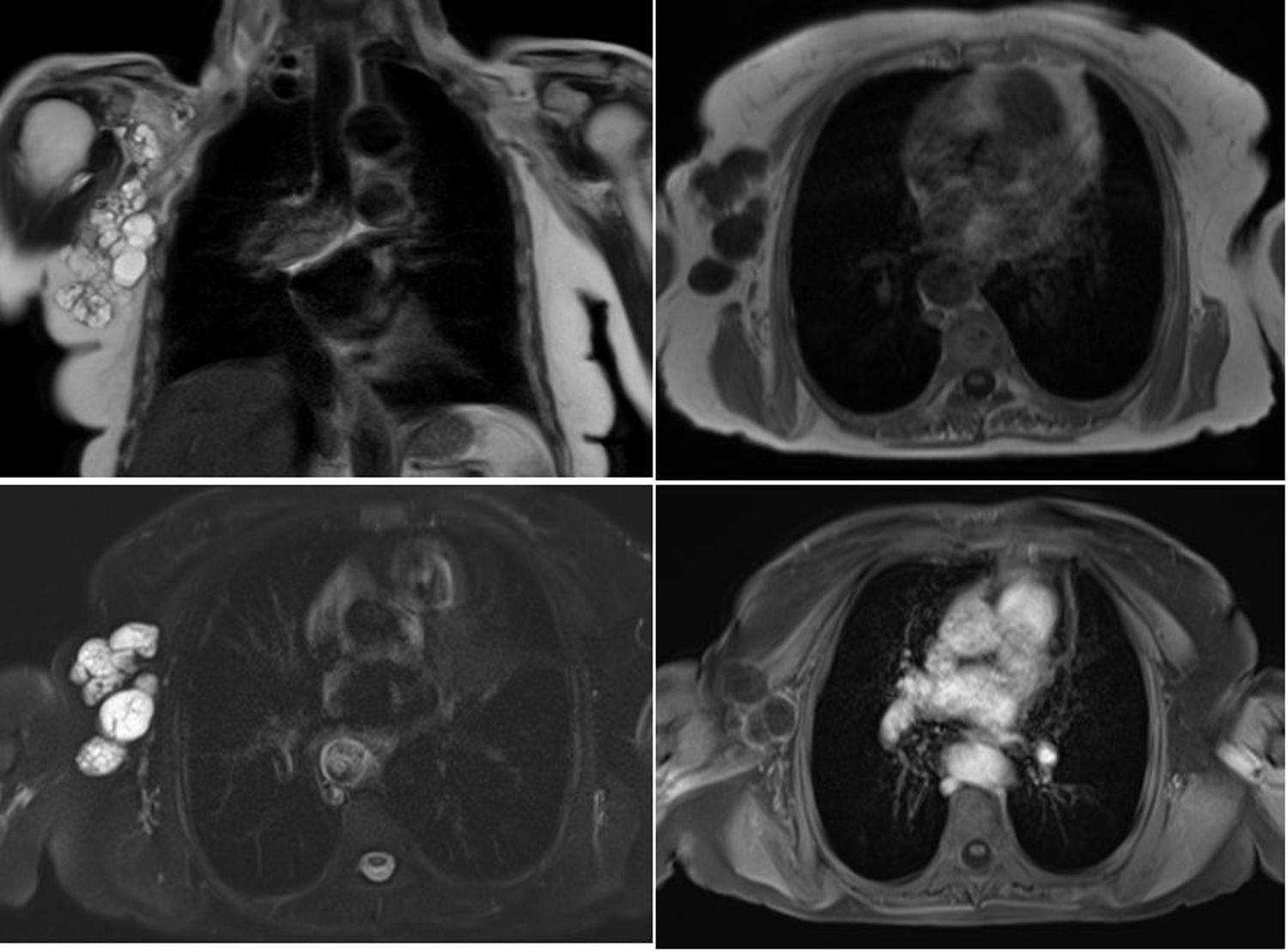
A 77-year-old female presented to our clinic with a mass in the right axilla. Coronal (Panel A) and axial (Panel B) T2-weighted MR images reveal a multiseptated cystic lesion solely in the right axillary fossa. Axial pre- (Panel C) and post-contrast (Panel D) T1-weighted MR images show multiple cysts with peripheral enhancement of their walls following gadolinium administration.

Fig. 1

Although extremely rare, clinicians should include hydatid disease in the differential diagnosis of palpable axillary mass lesions in endemic regions.

Copyright © 2020. AEC
Download PDF
Article options
Tools