


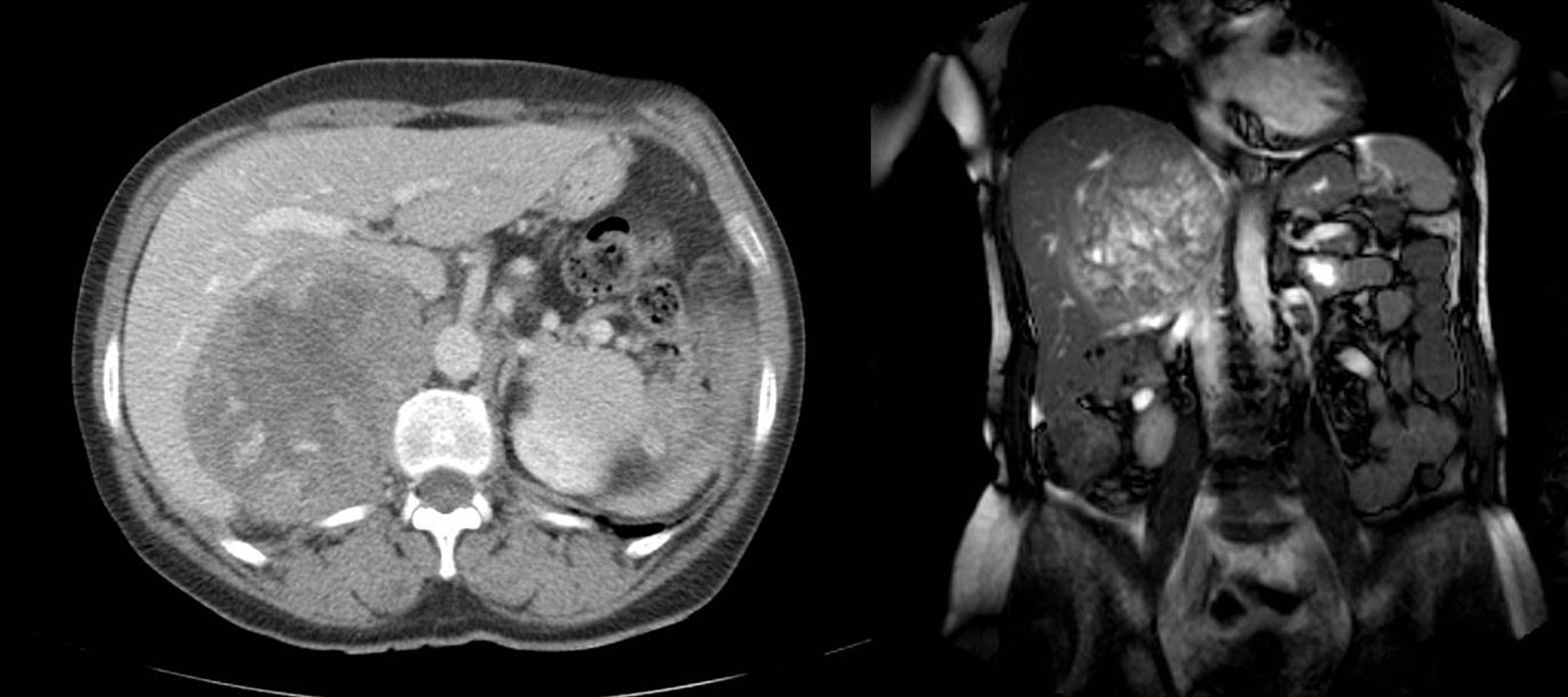
A 64-year-old patient with no prior medical history of interest presented with abdominal pain and a mass effect in the upper right quadrant that had been progressing for the previous 3 months, with no other symptoms. CT and MRI studies (Fig. 1) detected a voluminous retroperitoneal mass (approximately 12×12×8cm) that encompassed the right adrenal gland, pressing on and deforming the hepatic outline, which compressed and displaced the inferior vena cava, although no infiltration was seen. The adrenal function was analyzed, and no alterations were found.
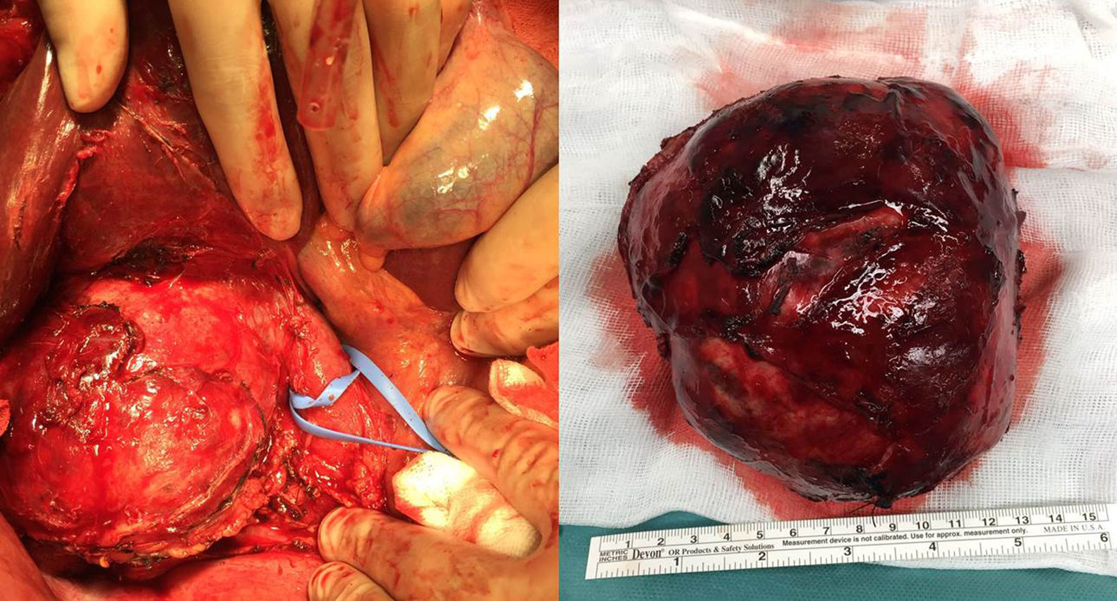
With a diagnostic suspicion of adrenal neoplasm, the patient was treated surgically (Fig. 2), and the pathology study reported giant adrenal cortical carcinoma.
Please cite this article as: Estévez Fernández S, Artime Rial M, Domínguez Comesaña E, Sánchez Santos R. Carcinoma cortical adrenal gigante. Cir Esp. 2017;95:542.
