


Taphonomy helps to understand the issues related to changes of the cadaveric remains in the frame of palaeontology and archaeology as well as in the frame of forensic anthropology. The first objective of the experimental project Taphos-m was to generate a corpus of information on taphonomy to know what taphonomic agents and process could be responsible for the observable effects in field.
Materials and methodsThe cadaveric state of Sus scrofa domestica remains and the spatial distribution of the anatomical elements has been described. In the case of skeletonisation, the state of the cortical surface and fragmentation of the bones was evaluated too. Also the pathological and histological analysis has been observed.
ResultsThe animal remains buried in the stone tomb were in dried state, while the remains buried in the tile tomb were skeletonised. There were differences in the observable taphonomic effects, particularly in the spatial distribution of the anatomical elements. The lesion in the leg of one animal could be responsible of the maintenance of anatomic articulation.
ConclusionsMeteorological data during inhumation and the tomb characteristics are variables that determine the evolution and condition of the remains, but they are not the only ones: the pathological lesions may involve differences in the spatial distribution of the bones and anatomical articulations.
La tafonomía ayuda a entender las cuestiones relacionadas con las modificaciones post mortem de los restos cadavéricos en los campos de la paleontología, la arqueología y la antropología forense. Por ello, el objetivo principal del proyecto experimental Taphos-m es generar un corpus en tafonomía que permita comprender qué agentes y procesos tafonómicos son los responsables de los efectos observados en diferentes contextos.
Material y métodosPasados 3 años y medio desde su inhumación, se ha valorado el estado cadavérico de 2 cuerpos de Sus scrofa domestica enterrados en 2 estructuras vacías de características constructivas distintas, así como la distribución espacial que presentaban los elementos anatómicos. En caso de esqueletización, también se describe el estado de la superficie cortical de los huesos y la eventual fragmentación ósea. También se han llevado a cabo analíticas complementarias, como análisis histológico y patológico.
ResultadosLos restos del animal enterrado en la tumba de piedra se encontraron en estado desecado, mientras que los restos inhumados en la tumba de tejas planas estaban prácticamente esqueletizados. Se observaron diferencias en el análisis de los efectos tafonómicos, sobre todo en relación con la distribución espacial de los elementos anatómicos, vinculados con la presencia de sedimento y el estado cadavérico. La lesión que presentaba uno de los animales en la extremidad trasera podría influir en el mantenimiento de la articulación anatómica.
ConclusionesLos datos meteorológicos del momento de la inhumación y las características de la tumba son variables que determinan la evolución y el estado cadavérico de los restos, pero no son las únicas, ya que las lesiones pueden suponer diferencias en la distribución espacial de los restos óseos y articulaciones anatómicas.
Taphonomy (from the Greek taphos, meaning “burial”, and nomos, meaning “law”) is the study of the chemical, physical, biological and geological processes occurring in corpses from the moment of death until their recovery.1 Taphonomic studies are therefore intended to be a source of useful information for understanding the events that occurred and the postmortem interval in the field of forensic medicine and anthropology.2–4 Similarly, the analysis of taphonomic effects can help to understand the funerary practices and treatment of death in ancient populations.5,6
Currently, there are very few palaeontological and anthropological studies that link taphonomic effects observed on site with known agents and processes.7–9 Human decomposition has been studied using limited experiences such as those of the Forensic Anthropology Centre at the University of Tennessee, Knoxville.10 The difficulties in undertaking human experimentation have boosted the use of animal models, especially the species Sus scrofa domestica (Linnaeus 1758) for its similarity to the human body with regard to amount of hair, torso size, intestinal flora, eating habits, and decomposition processes.11,12
The pilot project Taphos-m13,14 was initiated in 2011, and it is based on the analysis of the decomposition of domestic pigs (S. scrofa domestica). Facilities include 26 burials controlled by the type of funerary structure, position and body characteristics, meteorological data, and elements specific to the remains. The project aims at testing whether individuals with similar characteristics that are buried under the same conditions, but in different funerary structures (structures infilled with soil vs empty space structures), show skeletal differences at the time of exhumation. Apart from other minor structural differences in the tombs, it seems that the main differences should be in relation to whether the structures are infilled or empty space. In infilled structures, corpses are buried in direct contact with sediment, so that as it decomposes soft tissue, it is replaced by soil, keeping skeletal elements well articulated. Empty space structures, however, contain corpses that are not covered with sediment, so that during the decomposition, the corpse's bones are displaced by gravity to the base of the structure, losing the original anatomical connection.3
Three and a half years after burying the corpses in the Taphos-m pilot enclosure, we opened 2 empty space structures (5-26 and 6-08) in order to review the status of the corpses and assess the degree in which their joints were held together. The opening of these 2 tombs aimed at distinguishing whether: (a) preservation and articulation differences exist in a corpse based on whether the type of empty structure is more or less sealed,3,15 and (b) the presence of lesions affecting the articulation or dislocation of anatomical areas is involved.16,17
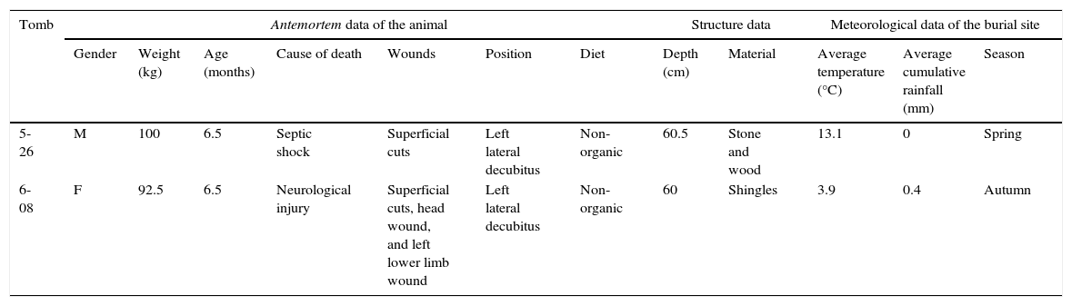
Material and methodsFor this study, 2 tombs were chosen that allowed for a comparison and evaluation of very specific variables based on to the animal's characteristics (gender, weight, cause of death, and presence of wounds) and the funerary structure that housed them (material) (Table 1). Animal 5-26 was a young (6.5 months) male pig, whose diet was non-organic. He died of septic shock, and at the time of burial he had wounds on the head and tail. He was buried in an empty structure with stone-panelled walls, original floor, and wooden roof. Animal 6-08, whose age and diet were the same as animal 5-26, was a female pig that died from a neurological injury. In this case, the animal, which had wounds on the head and left hind limb, was buried in an empty structure made of shingles (Fig. 1). Both structures are oriented North-South and are contiguous, with tomb 6-08 being closer to the hillside just East of the facilities. The stone structure with a wooden roof (5-26) has been used as a control to check the progress of the remains over the years, having been opened a total of 10 times since 2012.
Information on animals 5-26 and 6-08 obtained at the time of burial: antemortem data, data on burial structures, and meteorological data of the burial sites.
| Tomb | Antemortem data of the animal | Structure data | Meteorological data of the burial site | |||||||||
|---|---|---|---|---|---|---|---|---|---|---|---|---|
| Gender | Weight (kg) | Age (months) | Cause of death | Wounds | Position | Diet | Depth (cm) | Material | Average temperature (°C) | Average cumulative rainfall (mm) | Season | |
| 5-26 | M | 100 | 6.5 | Septic shock | Superficial cuts | Left lateral decubitus | Non-organic | 60.5 | Stone and wood | 13.1 | 0 | Spring |
| 6-08 | F | 92.5 | 6.5 | Neurological injury | Superficial cuts, head wound, and left lower limb wound | Left lateral decubitus | Non-organic | 60 | Shingles | 3.9 | 0.4 | Autumn |
 details of the lower third of the body; the circle indicates the wound in the left hind limb at the time of burial; (b) details of the lesion.")
The study of the remains in tombs 5-26 and 6-08 was based on: (a) the description of the cadaveric state, and (b) the analysis of the observed taphonomic effects. Although the process of decomposition of a corpse in nature is a continuous process,11 different cadaveric states can be described.4 For the taphonomic analysis, macroscopic changes affecting bone record in 3 different areas were observed: changes in the original distribution or position of the skeletal elements (from tight articulation to disarticulation),3,12,15 changes at the cortical surface of the bone, and bone fragmentation.12,18
During excavation, desiccated tissue samples were taken from animal 5-26 for histological analysis. The methodology used for laboratory analysis was based on hydration with Sanderson solution, formalin fixation (10%), paraffin sections, and haematoxylin-eosin staining.19 Finally, a study of bone lesions was also performed.
ResultsRegarding tomb 5-26, holes and cracks were observed in its outer perimeter, being more striking in the southern part of the tomb corresponding to the caudal part of the animal. The animal's body was found partially covered with sediment, soil, gravel, and small stones that had entered through the roof, affecting the observation of the lower third of the body. Desiccated soft tissue was visible, which virtually covered the entire exposed body (Fig. 2a). Under the desiccated tissue, the remains were in the skeletal reduction stage (Fig. 2b). No desiccated tissue was found in the hind limbs, which were covered with sediment. Instead, they were in a total skeletal reduction stage (Fig. 2c). These external characteristics correspond to a desiccated/mummified corpse state. Histological analysis confirmed that the desiccated tissue was compatible with skin (Fig. 2d).
 desiccated soft tissue covering the part of the body exposed; (b) skeletonisation of the remains under the desiccated tissue; (c) total skeletonisation of the rear limb infilled with soil (the circle indicates the presence of hooves); and (d) histological image (haematoxylin–eosin staining, magnified ×10) of the mummified tissue in which keratinised epidermis (1) and dermis (2) can be seen.")
Cadaveric state of animal 5-26: (a) desiccated soft tissue covering the part of the body exposed; (b) skeletonisation of the remains under the desiccated tissue; (c) total skeletonisation of the rear limb infilled with soil (the circle indicates the presence of hooves); and (d) histological image (haematoxylin–eosin staining, magnified ×10) of the mummified tissue in which keratinised epidermis (1) and dermis (2) can be seen.
The bones of animal 5-26 were well preserved, with no changes to the cortical surface, fragmentation, or loss of bone substance. Their colour was dark brown and had the characteristic appearance of a non-adult skeleton (porosity and growth striations). Overall, the skeleton's articulations were tight, except for some anatomical areas that had a looser articulation (Fig. 3): cervical vertebrae were not tight, whereas the thoracic and lumbar vertebrae showed a high degree of articulation. The ribs on both sides had lost their initial anatomical position, having a loose articulation (Fig. 3). The epiphysis of the long bones and the vertebral bodies were not fused, although they maintained their anatomical position (Fig. 3). The hind limb, which was covered with sediment, closely maintained its original position (Figs. 2c and 3).

The external cracks in tomb 6-08, in contrast, were not as obvious as in structure 5-26, even though this structure was also not infilled. The animal's body was found surrounded by a mass with a thickness of 1cm, formed by putrilage, mud, and puparia masses. All bones were visible, except for the skull, since the head was found partially mummified (Fig. 4a). Small traces of soft tissue were also found in the right forelimb and lower back (Fig. 4b), while remains of the hooves were preserved in the hind limbs. The remains of this animal correspond to a cadaveric state, which includes the skeletal phase and tissue mummification.
 virtually total skeletonisation of remains and desiccated skull tissue; (b) hair remains in the lumbar area (arrow).")
Overall, no external taphonomic effects were observed, with bones being darker and homogeneous in texture, and with no changes that deteriorate the cortical surface, except for the typical porosity of juvenile bones. The skeleton maintained all its articulations loosely, with the lower back and left forelimb being the only anatomical regions where articulations were tight (Fig. 5a). The most remarkable disarticulation was observed in the left hind limb (Fig. 5b), the anatomical part where the animal had a wound (Fig. 1). This left limb injury comprised an abnormal proliferation of the calcaneus.
Discussion tight articulation of the left forelimb (arrow); (b) disarticulation of the left hind limb (arrow).")
In some cases, the absence of information and data on the context and environment in pathology and anthropology studies renders the interpretation of observable taphonomic effects in funeral deposits difficult.9,18 As a result, the Taphos-m pilot project aims at providing data to help understand taphonomic effects observed in an experimental space, and interpret the characteristics and causes of cadaveric state and the disposition of burial sites.13,14 This study provides the first data of the pilot project, based on the opening of empty space funerary structures 5-26 and 6-08.
The funerary structures that form the Taphos-m project have changed and deteriorated. It is evident that since the time of burial, the structures located in the East, along the hillside, have been the most affected, because the landslide has almost eroded their location. The presence of soil inside the stone grave with a wooden roof (No 5-26) must be related to cracks and holes found at the outer limits of the southern part of the tomb, as well as the fact that this structure is covered by wood, a porous material. In this regard, the deterioration of tomb 5-26 allowed for the entry of non-original sediment, which probably penetrated the structure slowly over the 3years since its closure. The numerous occasions when the tomb was opened to observe the state of the remains have also influenced the entry of sediment. That is why the southern part of the tomb (the most cracked surface area) was affected and the animal's hind limbs were found covered in this sediment. The results show that the shingle structure (No 6-08) was more resistant to the entry of sediment, despite being closer to the mountain, which was gradually collapsing. Still, the presence of non-original, fine, and homogeneous sediment, covering the entire inner base of tomb 6-08, was evident. Nonetheless, this sediment did not hinder the observation of the animal's remains.
The valuation of macroscopic taphonomic effects showed that the bones had a dark colour, which is seen in remains of corpses that have been dead less than 5 years and buried in empty spaces.20 This being a case of 2 young animals, the epiphysis of the bones were not fused, and bones showed a porosity and striation typical of bones of juvenile individuals.20 The cadaveric state of the remains was, however, different in each case: animal 5-26 was found desiccated, whereas animal 6-08 was found at the stage of total skeletal reduction.4 Specifically, the anatomical areas of animal 5-26 that came into contact with the soil base were fully skeletonised, as were the hind limbs, which were completely covered with sediment that subsequently entered the tomb. In this sense, it seems that skeletonisation progresses faster and tissue desiccation is not as usual when the corpse is in direct contact with sediment.2 Regarding the preservation of each animal's corpse, these differences could be related, besides the type of structure that houses them, to climatological phenomena occurring at death and burial21,22 (Table 1): the spring burial, with low humidity and high temperatures (No 5-26), seems to have facilitated mummification, whereas the autumn burial, in colder conditions and in an airtight structure—without air-circulation—(No 6-08), facilitated the total skeletonisation of the corpse. As has been described by other authors,2,3 the cause of death and gender may also influence the cadaveric state of the remains, although these factors were not considered in the 2 cases presented here. Future openings of the other funerary structures of the Taphos-m pilot enclosure could confirm or disprove these hypotheses.
The taphonomic effects observed are also related to differences in the spatial distribution of anatomical elements in the tomb.3,12,15 Overall, the corpse of animal 5-26 was tightly articulated, probably thanks to soft tissue preservation, which helped to preserve anatomical articulations. The hind limb, which was covered with sediment deposited at a later date, presented with greater articulation. The differential preservation of the corpses between the structure filled with soil and the empty space structure is evident, and is directly related to the presence or absence of sediment in direct contact with the remains.2,15
The articulations of animal 6-08 were looser, except for the left hind limb which, despite being in contact with sediment, showed the greatest disarticulation, especially at the calcaneus and talus bone. This disarticulation is related to the antemortem pathological lesion presented by the animal on that limb, suggesting that the presence of a wound at the time of burial was a means of access for cadaveric fauna, a factor that manifests as the disarticulation of the left hind limb.2,16,17
ConclusionsThe opening of 2 empty structures at the Taphos-m pilot enclosure allowed us to observe that the taphonomic differences of 2 empty space burials are related to the biological characteristics of the animal (gender, age, and cause of death), meteorological data, and tomb-related characteristics, which determine the entry of sediment and organisms. The presence of lesions significantly influences the process of decomposition of the corpse and has an effect on its state of articulation. This experience suggests the great interplay of variables that can intervene in the cadaveric state of the remains, and the importance of considering them when interpreting the origin and condition of forensic remains.
Conflicts of interestThe authors declare that they have no conflicts of interest.
The members of Grup de Recerques Terres de Ponent and of Institut d’Estudis Ilerdencs, who provided the necessary materials for this study. Drs Ferran Jiménez and Albert Isidro for performing the bone histology and pathology analyses, respectively.
Please cite this article as: Gutiérrez A, Nociarová D, Malgosa A, Armentano N. Comparación de los efectos tafonómicos observados en dos estructuras funerarias de espacio vacío. Rev Esp Med Legal. 2016;42:98–104.
Some information for this manuscript was presented at the Seventh Meeting of the Association Espanola Anthropology and Odontology Forensic, on 6 and 7 November 2015 in Toledo.
articles