Buscar en
Radiología (English Edition)
Toda la web
Inicio Radiología (English Edition) Evaluation of extramedullary plasmacytoma of the larynx with radiologic and hist...
Journal Information
Share
Share
Download PDF
More article options
Visits
102
Brief report
Evaluation of extramedullary plasmacytoma of the larynx with radiologic and histopathological findings
Evaluación del plasmocitoma extramedular de laringe con hallazgos radiológicos e histopatológicos
Visits
102
A. Tanrivermis Sayita,
Corresponding author
, M. Elmalia, S. Günb
a Departamento de Radiología, Facultad de Medicina de la Universidad Ondokuz Mayis, Samsun, Turkey
b Departamento de Anatomía Patológica, Facultad de Medicina de la Universidad Ondokuz Mayis, Samsun, Turkey
This item has received
Received 03 July 2019. Accepted 16 July 2020
Article information
Abstract
Full Text
Bibliography
Download PDF
Statistics
Figures (3)
Show moreShow less
Abstract

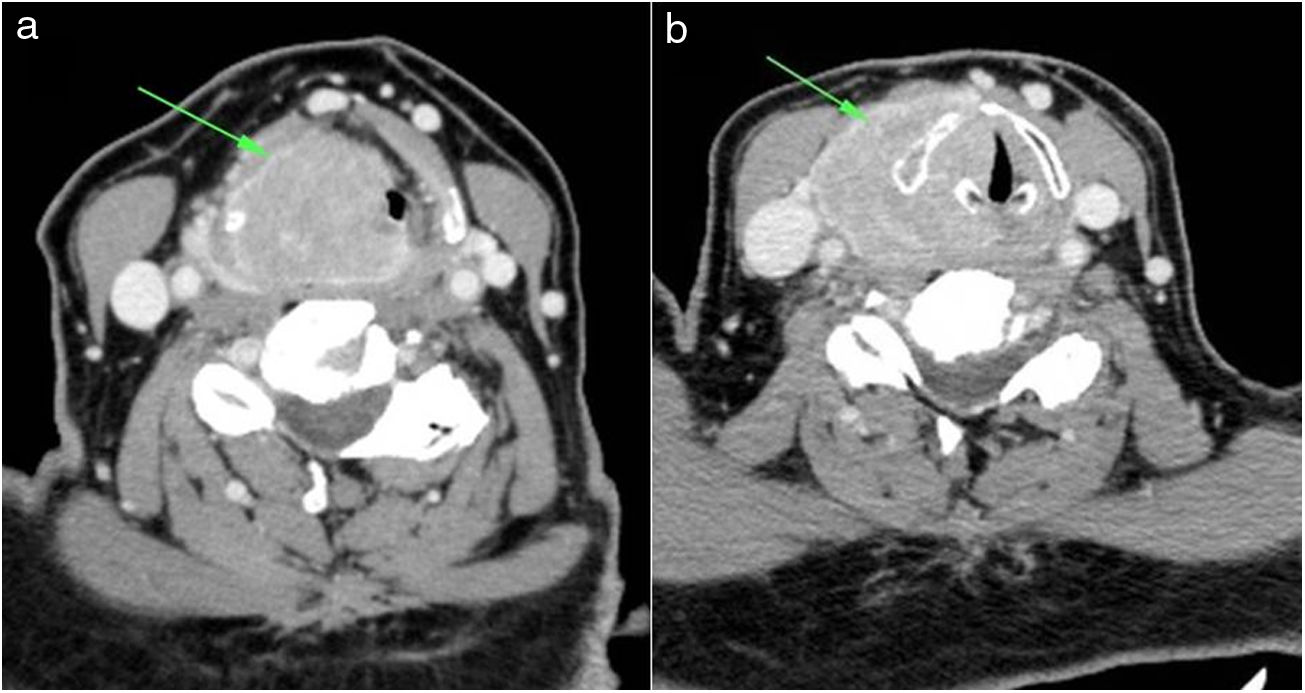
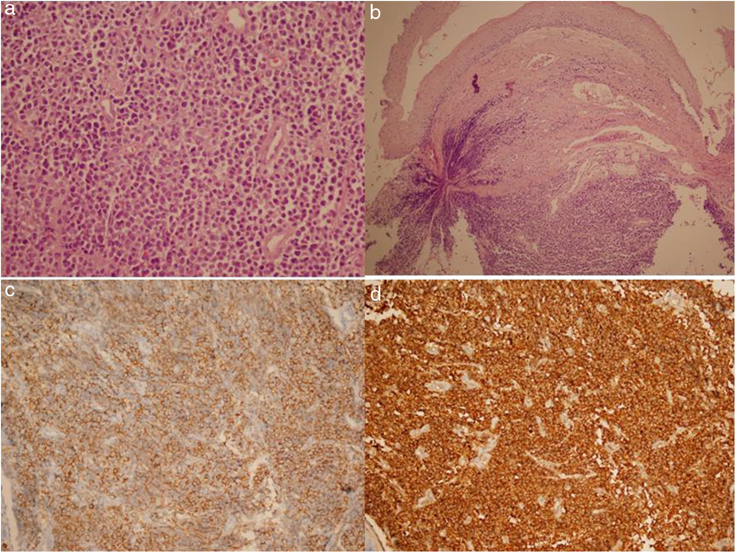
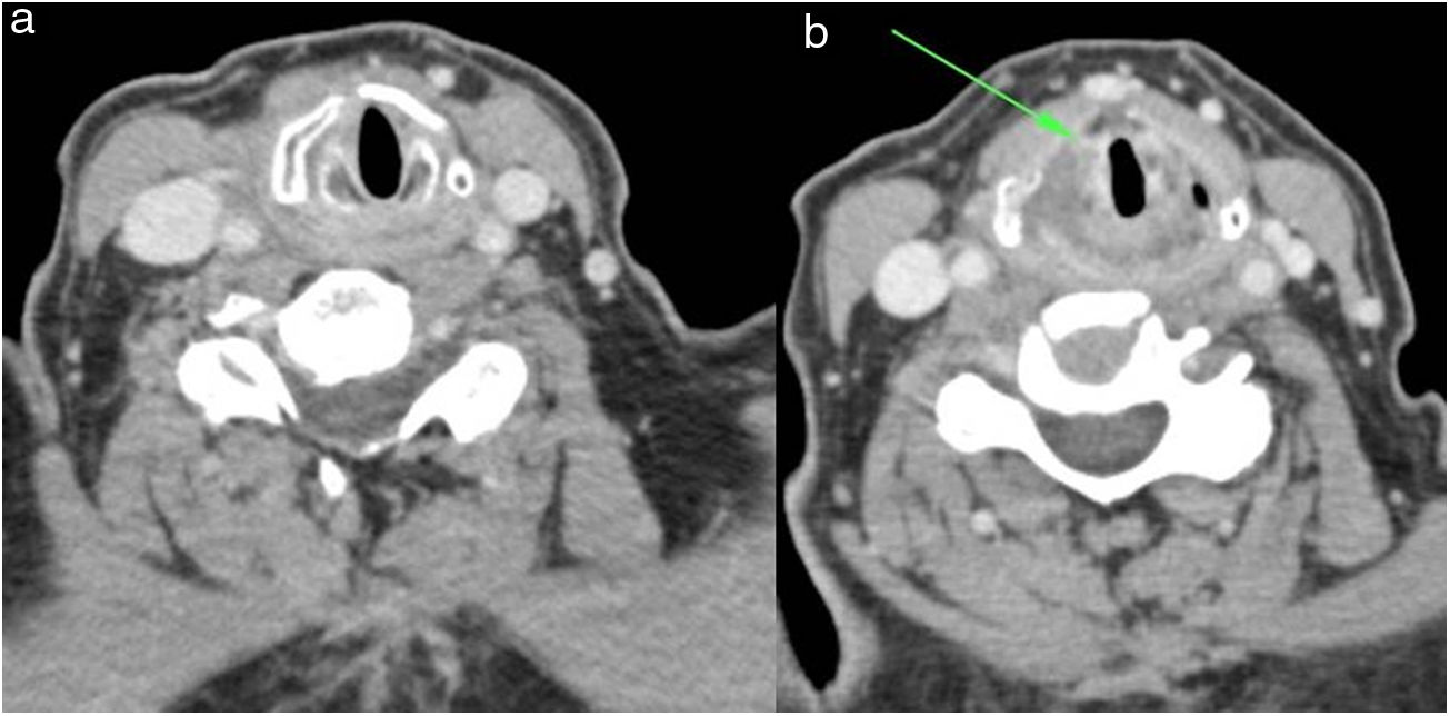
Extramedullary plasmacytoma (EMP) is a plasma cell neoplasm of soft tissue without bone marrow involvement or other systemic characteristics of multiple myeloma. Approximately 80%–90% of EMPs involve the head and neck region, especially in the nasal cavity, paranasal sinuses, tonsillar fossa, and oral cavity. An EMP of the larynx is extremely rare and is a locally destructive lesion without systemic spread. Clinical features vary depending on the tumor location. A diagnosis is established by histopathology, immunohistochemistry, and a systemic survey to exclude systemic plasma cell proliferative diseases. Extramedullary plasmacytomas are highly radiosensitive and radiotherapy is therefore used as a treatment. In this study, we report on a rare case of EMP of the larynx evaluated with computed tomography and present histopathologic findings for a 74-year-old female patient.

Keywords:
Computed tomography
Extramedullary
Solitary plasmacytoma
Larynx
Resumen

El plasmocitoma extramedular (PEM) es una neoplasia de células plasmáticas del tejido blando sin afectación de la médula ósea ni otras características sistémicas de mieloma múltiple. Aproximadamente el 80%–90% de los PEM afectan a la región de la cabeza y el cuello, especialmente a la cavidad nasal, los senos paranasales, la fosa amigdalina y la cavidad oral. Los casos de PEM de laringe son extremadamente raros y se trata de una lesión destructiva a nivel local sin propagación sistémica. Las características clínicas varían en función de la localización del tumor. El diagnóstico se establece mediante histopatología, inmunohistoquímica y un estudio sistémico para excluir las enfermedades sistémicas de proliferación de células plasmáticas. Los plasmocitomas extramedulares son altamente sensibles a la radiación, por lo que se utiliza la radioterapia como tratamiento. En este estudio, notificamos un caso raro de PEM de laringe evaluado con tomografía computarizada y presentamos los resultados histopatológicos de una paciente de 74 años.

Palabras clave:
Tomografía computarizada
Extramedular
Plasmocitoma solitario
Laringe

Article

These are the options to access the full texts of the publication Radiología (English Edition)
Subscriber
Subscriber

If you already have your login data, please click here .

If you have forgotten your password you can you can recover it by clicking here and selecting the option “I have forgotten my password”
Purchase
Purchase article

Purchasing article the PDF version will be downloaded

Price 19.34 €

Purchase now
Contact
Phone for subscriptions and reporting of errors
From Monday to Friday from 9 a.m. to 6 p.m. (GMT + 1) except for the months of July and August which will be from 9 a.m. to 3 p.m.
Calls from Spain
932 415 960
Calls from outside Spain
+34 932 415 960
E-mail
Article options
Tools
es en pt

¿Es usted profesional sanitario apto para prescribir o dispensar medicamentos?

Are you a health professional able to prescribe or dispense drugs?

Você é um profissional de saúde habilitado a prescrever ou dispensar medicamentos