


To explore whether children and adolescents with attention deficit/hyperactivity disorder (ADHD) have altered the functional connectivity between the executive control network and the default mode network.
MethodsExploratory study of a diagnostic test, prospective, case and control design. A total of 56 participants were recruited consecutively (29 inattentive or combined ADHD subtype and 27 controls) between 7 and 16 years old, male, right dominance. DSM-5 was applied as reference test and a battery of neuropsychological tests to confirm the diagnosis and assess comorbidities. Resting state functional magnetic resonance imaging was performed as an index test. The application and evaluation of the tests was blind. The brain regions were chosen a priori and the region of interest technique was used. The functional connectivity of the anterior cingulate cortex (ACC) was evaluated with: the precuneus (P), the posterior cingulate cortex (PCC) and the dorsomedial prefrontal cortex (DMPC).
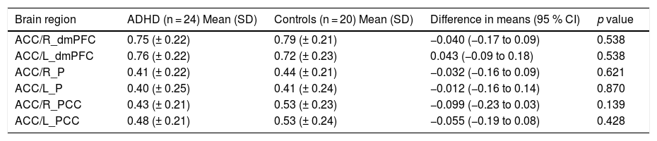
ResultsThe functional connectivity in each of the associations evaluated in the patients with ADHD compared with the controls were: P_D = 0.41 vs 0.44; CCP_D = 0.43 vs 0.53; CPDM_D = 0.75 vs. 0.79; P_I = 0.40 vs 0.41; CCP_I = 0.48 vs 0.53; CPDM_I = 0.76 vs. 0.72). D: right side I: left side. Value of p > 0.05.
ConclusionCerebral functional connectivity at rest is lower in ADHD patients when compared with healthy controls, however, the difference was not statistically significant.
Explorar si los niños y adolescentes con trastorno por déficit de atención e hiperactividad (TDAH) tienen alterada la conectividad funcional entre la red de control ejecutivo y la red neuronal por defecto.
Material y métodosEstudio exploratorio de prueba diagnóstica, prospectivo, con diseño de casos y controles. Se reclutaron consecutivamente 56 participantes (29 con TDAH de tipo inatento o combinado y 27 controles) entre 7 y 16 años, de sexo masculino, dominancia derecha. Se aplicaron los criterios diagnósticos del DSM-5 como prueba de referencia y una batería de pruebas neuropsicológicas para confirmar el diagnóstico y evaluar comorbilidades. Se les realizó resonancia magnética funcional de reposo como prueba índice. La aplicación y evaluación de las pruebas fue ciega. Las regiones cerebrales se escogieron a priori y se usó técnica de región de interés. Se evaluó la conectividad funcional de la corteza del cíngulo anterior (CCA) con el precuneus (P), la corteza del cíngulo posterior (CCP) y la corteza prefrontal dorsomedial (CPDM).
ResultadosLas conectividades funcionales en cada una de las asociaciones evaluadas en los pacientes con TDAH comparado con los controles fueron: P_D = 0,41 vs. 0,44; CCP_D = 0,43 vs. 0,53; CPDM_D = 0,75 vs. 0,79; P_I = 0,40 vs. 0,41; CCP_I = 0,48 vs. 0,53; CPDM_I = 0,76 vs. 0,72). D: lado derecho I: lado izquierdo. Valor de p > 0,05.
ConclusiónLa conectividad funcional cerebral en estado de reposo es menor en los pacientes con TDAH cuando se compara con controles sanos; sin embargo, la diferencia no fue estadísticamente significativa.

