


Cerebral small vessel ischemic disease (SVID) as a common age-related morbidity is the key mechanism of vascular cognitive impairment (VCI). This study uses Cerebral blood flow (CBF) measured by pseudo-continuous ASL MRI in SVID patients with and without cognitive impairment to differentiate VCI from normal aging.
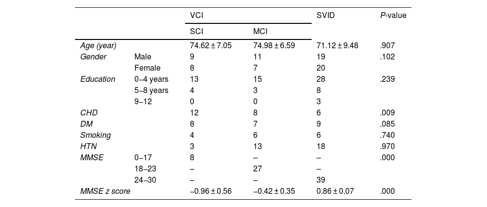
Materials and methodsIn this cross-sectional study, 74 SVID patients, including 35 with diagnosed VCI and 39 without cognitive impairment (control) underwent pCASL-MRI in the resting state. ROI-based approach pre-processing, denoising techniques, and correction for partial volume effects were performed. Regional CBF was compared between severe cognitive impairment (SCI), mild cognitive impairment (MCI), and SVID patients without cognitive impairment.
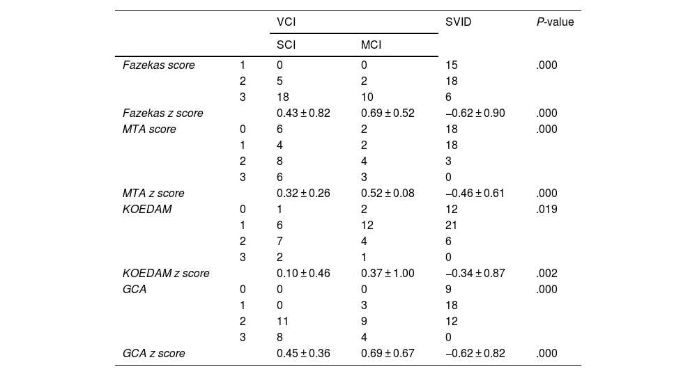
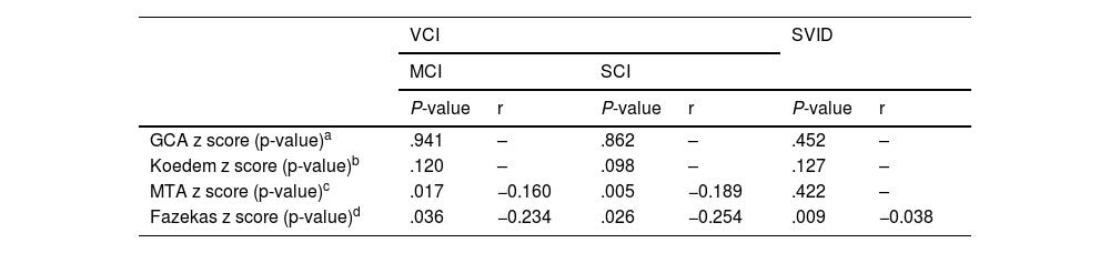
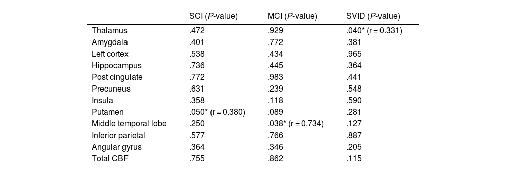
ResultsTotal and regional CBF values in the thalamus, left cortex, hippocampus, post cingulate cortex, precuneus, insula, putamen, and middle temporal lobe was lower in VCI compared to SVID, also in SCI compared MCI group. There was a linear correlation between the Mini-Mental State Examination (MMSE) z score and CBF in the thalamus region in SVID participants and between the MMSE z score and CBF in the medial temporal region in MCI participants. The medial temporal atrophy )MTA( z score was significantly correlated with CBF values in the hippocampus and medial temporal regions in SCI and MCI also a significant correlation was seen between total CBF and Fazekas score.
ConclusionDue to the growing prevalence of dementia and the role of CBF as a predictive biomarker, ASL-MRI as a non-invasive method can be easily added to diagnostic tools of cognitive impairment in individuals with SVID to recognize the initiation of vascular cognitive impairment.
La enfermedad isquémica cerebral de pequeño vaso (EIPV), morbilidad común relacionada con la edad, es el mecanismo clave del deterioro cognitivo vascular (DCV). Este estudio utiliza el flujo sanguíneo cerebral (FSC) medido mediante RM-ASL pseudocontinua en pacientes con EIPV con y sin deterioro cognitivo para diferenciar el DCV del envejecimiento normal.
Materiales y métodosEn este estudio transversal, 74 pacientes con EIPV, incluidos 35 con DCV diagnosticado y 39 sin deterioro cognitivo (control) se sometieron a una RM-pCASL en reposo. Se llevaron a cabo un preprocesamiento basado en ROI, técnicas de eliminación de ruido y corrección de los efectos de volumen parcial. Se comparó el FSC regional entre pacientes con deterioro cognitivo grave (DCG), deterioro cognitivo leve (DCL) y EIPV sin deterioro cognitivo.
ResultadosLos valores totales y regionales del FSC en el tálamo, la corteza izquierda, el hipocampo, la corteza cingulada posterior, la precuña, la ínsula, el putamen y el lóbulo temporal medio fueron más bajos en el grupo DCV en comparación con el de EIPV, y también en el grupo DCG en comparación con el de DCL. Se observó una correlación lineal entre la puntuación z de la minievaluación del estado mental (Mini-Mental State Examination, MMSE) y el FSC en la región del tálamo en los participantes con EIPV y entre la puntuación z del MMSE y el FSC en la región temporal medial en los participantes con DCL. La puntuación z de la atrofia temporal medial (ATM) se correlacionó significativamente con los valores del FSC en el hipocampo y las regiones temporales mediales en el DCG y el DCL. También se observó una correlación significativa entre el FSC total y la puntuación de Fazekas.
ConclusiónDebido a la creciente prevalencia de la demencia y al papel del FSC como biomarcador predictivo, la RM-ASL como método no invasivo puede añadirse fácilmente a las herramientas de diagnóstico del deterioro cognitivo en individuos con EIPV para reconocer el inicio del deterioro cognitivo vascular.

