Buscar en
Radiología (English Edition)
Toda la web
Inicio Radiología (English Edition) Imaging findings in neurocysticercosis
Journal Information
Vol. 55. Issue 2.
Pages 130-141 (March - April 2013)
Share
Share
Download PDF
More article options
Visits
9282
Vol. 55. Issue 2.
Pages 130-141 (March - April 2013)
Update in Radiology
Imaging findings in neurocysticercosis
Neurocisticercosis. Hallazgos radiológicos
Visits
9282
S. Sarria Estrada
Corresponding author
ssarria@idi-cat.org

Corresponding author.
, L. Frascheri Verzelli, S. Siurana Montilva, C. Auger Acosta, A. Rovira Cañellas
Unitat de Ressonància Magnètica (IDI), Servei de Radiologia, Hospital Universitari Vall d’Hebron, Barcelona, Spain
This item has received
Article information
Abstract
Full Text
Bibliography
Download PDF
Statistics
Figures (11)
Show moreShow less
Tables (1)
Table 1. Stages of neurocysticercosis.
Abstract

Neurocysticercosis, caused by the larvae of Taenia solium, is the parasitic infection that most commonly involves the central nervous system in humans. Neurocysticercosis is endemic in practically all developing countries, and owing to globalization and immigration it is becoming more common in developed countries like those in western Europe.

The most common clinical manifestations are epilepsy, focal neurologic signs, and intracranial hypertension.

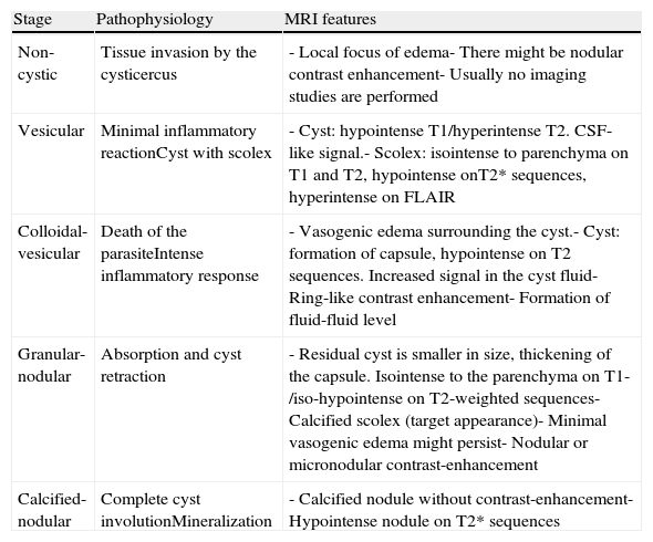
The imaging findings depend on the larval stage of Taenia solium, on the number and location of the parasites (parenchymal, subarachnoid, or intraventricular), as well as on the host's immune response (edema, gliosis, and arachnoiditis) and on the development of secondary lesions (arteritis, infarcts, or hydrocephalus).

The diagnosis of this parasitosis must be established on the basis of the clinical and radiological findings, especially in the appropriate epidemiological context, with the help of serological tests.

Keywords:
Taenia solium
Cysticercosis
Neurocysticercosis
Computed tomography
Magnetic resonance imaging
Resumen

La neurocisticercosis es una parasitosis humana causada por las larvas de la Taenia solium, que es la que con mayor frecuencia afecta el sistema nervioso central. Esta infección es endémica en prácticamente todos los países en vías de desarrollo, pero debido a la globalización y a las migraciones humanas su frecuencia ha aumentado en países desarrollados como los de Europa Occidental.

Las manifestaciones clínicas más frecuentes son la epilepsia, signos neurológicos focales e hipertensión intracraneal.

Los hallazgos radiológicos dependen del estadio larvario de la Taenia solium, número y localización de los parásitos (parenquimatosa, subaracnoidea e intraventricular), así como de la respuesta inmune del huésped (edema, gliosis, aracnoiditis) y del desarrollo de lesiones secundarias (arteritis, infartos o hidrocefalia).

El diagnóstico de esta parasitosis debe establecerse en función de los hallazgos clínicos y radiológicos, especialmente en un contexto epidemiológico adecuado, con apoyo de la serología.

Palabras clave:
Taenia solium
Cisticercosis
Neurocisticercosis
Tomografía computarizada
Imagen por resonancia magnética

Article

These are the options to access the full texts of the publication Radiología (English Edition)
Subscriber
Subscriber

If you already have your login data, please click here .

If you have forgotten your password you can you can recover it by clicking here and selecting the option “I have forgotten my password”
Purchase
Purchase article

Purchasing article the PDF version will be downloaded

Price 19.34 €

Purchase now
Contact
Phone for subscriptions and reporting of errors
From Monday to Friday from 9 a.m. to 6 p.m. (GMT + 1) except for the months of July and August which will be from 9 a.m. to 3 p.m.
Calls from Spain
932 415 960
Calls from outside Spain
+34 932 415 960
E-mail
Article options
Tools
es en pt

¿Es usted profesional sanitario apto para prescribir o dispensar medicamentos?

Are you a health professional able to prescribe or dispense drugs?

Você é um profissional de saúde habilitado a prescrever ou dispensar medicamentos

Quizás le interese:
10.1016/j.rxeng.2019.06.003
No mostrar más