


To qualitatively and quantitatively explore the spectral study of focal liver lesions, comparing it with the usual polychromatic assessment with single-energy computed tomography.
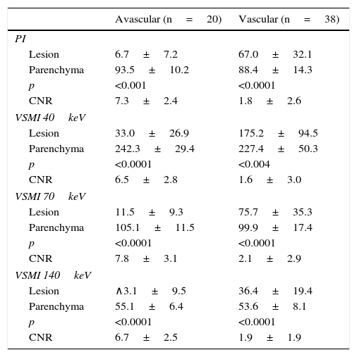
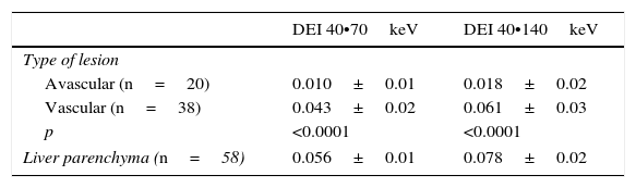
Material and methodsWe prospectively studied 50 patients with at least one focal liver lesion who were referred for abdominal multidetector computed tomography with intravenous contrast material. The portal phase was acquired with dual energy sources. The density of the lesions and of the surrounding liver parenchyma was measured both in the baseline polychromatic acquisition and in the posterior monochromatic reconstructions at 40keV, 70keV, and 140keV. Spectral curves were traced and the dual-energy indices and contrast-to-noise ratio were calculated. Lastly, the quality of the images and the detectability of the lesions were assessed qualitatively.
ResultsDensitometric differences between the different types of lesions (avascular and vascularized) and the liver were greater at low energy levels (left side of the spectral curve) than in the polychromatic evaluation. In the subjective assessment, the 40keV energy level had the greatest lesion detectability.
ConclusionsMonochromatic spectral study with dual-energy computed tomography provides better lesion detectability at 40keV compared to that provided by the ordinary polychromatic evaluation.
Explorar cualitativamente y cuantitativamente el estudio espectral de las lesiones focales hepáticas, comparándolo con la valoración policromática habitual de la tomografía de energía simple.
Mèc)todoEl presente estudio prospectivo incluyó 50 pacientes remitidos para la realización de tomografía computada multidetector abdominal con contraste intravenoso, que tuvieran al menos una lesión focal hepática. La fase portal fue adquirida con energía dual. Se realizaron mediciones densitomèc)tricas de las lesiones y del parèc)nquima hepático circundante, tanto en la adquisición policromática basal como en las posteriores reconstrucciones monocromáticas a 40, 70 y 140 keV. Se trazaron las curvas espectrales y se calcularon los índices de doble energía y la relación contraste-ruido. Por último, se hizo una valoración subjetiva de la calidad de las imágenes y la detectabilidad lesional.
ResultadosLas diferencias densitomèc)tricas entre los distintos tipos de lesiones (avasculares y vascularizadas) y el hígado fueron mayores a bajos niveles energèc)ticos (a la izquierda de la curva espectral) que en la evaluación policromática. En la valoración subjetiva, el nivel energèc)tico de 40keV presentó una mayor detectabilidad lesional.
ConclusionesEl estudio espectral monocromático mediante tomografía computada de energía dual provee una mayor detectabilidad lesional a 40keV en relación a la que se dispone con la valoración policromática habitual.

