metricas
Gastroenterología y Hepatología (English Edition) Implementation of esophageal endoscopic submucosal dissection in Spain: Results ...
Journal Information
Visits
381
Vol. 47. Issue 2.
Pages 119-129 (February 2024)
Original article
Full text access

Implementation of esophageal endoscopic submucosal dissection in Spain: Results from the nationwide registry

Implementación de la disección endoscópica submucosa esofágica en España: resultados del registro nacional
Visits
381
Enrique Rodríguez de Santiagoa,1,
Corresponding author
, Alberto Herreros-de-Tejadab,c,1, Eduardo Albénizd,1, Felipe Ramos Zabalae, Gloria Fernández-Esparrachf, Oscar Nogalesg, Pedro Rosónh, Beatriz Peñas Garcíaa, Hugo Uchimai,j, Álvaro Teránk, Joaquín Rodríguez Sánchezl,m, Diego de Frutosb, Sofía Parejo Carbonella, José Santiagob, José Díaz Tasendel, Charly Guarner Argenten, Pedro de María Pallaréso, Ana Amorósp, Daniel Barrancod, Daniel Álvarez de Castroa..., Raquel Muñoz Gonzálezi,j, José Carlos Marín-Gabriell, on behalf of the grupo GSEED de resección mucosa y endoscopia del 3° espacio de la Sociedad Española de Endoscopia Digestiva Ver más
a Servicio de Gastroenterología y Hepatología, Hospital Universitario Ramón y Cajal, Instituto Ramón y Cajal de Investigación Sanitaria (IRYCIS), Centro de Investigación Biomédica en Red en el Área temática de Enfermedades Hepáticas y Digestivas (CIBEREHD), Madrid, Spain
b Servicio de Digestivo, Hospital Universitario Puerta de Hierro, Instituto de Investigación Puerta de Hierro-Segovia de Arana (IDIPHISA), Majadahonda, Madrid, Spain
c Hospital La Luz, QuirónSalud, Madrid, Spain
d Unidad de Endoscopia, Servicio de Gastroenterología, Hospital Universitario de Navarra NavarraBiomed, Universidad Pública de Navarra (UPNA), IdisNA, Pamplona, Spain
e Servicio de Gastroenterología, Hospital Universitario HM Montepríncipe, Grupo HM hospitales, Boadilla del Monte, Madrid, Spain
f Sección de Endoscopia, Servicio de Gastroenterología, ICMDM, Hospital Clínic de Barcelona, Institut d’Investigacions Biomèdiques August Pi i Sunyer, CIBEREHD, Universidad de Barcelona, Barcelona, Spain
g Servicio de Gastroenterología, Hospital General Universitario Gregorio Marañón, Madrid, Spain
h Servicio de Aparato Digestivo, Hospital Vithas Xanit internacional Málaga, Málaga, Spain
i Hospital Universitari Germans Trias i Pujol, Badalona, Barcelona, Spain
j Centro Médico Teknon, Barcelona, Spain
k Servicio de Gastroenterología, Hospital Universitario Marqués de Valdecilla, Instituto de Investigación Valdecilla (IDIVAL), Santander, Spain
l Servicio de Medicina del Aparato Digestivo, Unidad de Endoscopias, Hospital Universitario 12 de Octubre, Instituto de Investigación «i+12», Madrid, Spain
m Servicio de Gastroenterología, Hospital General Universitario de Ciudad Real, Ciudad Real, Spain
n Servicio de Gastroenterología, Hospital de la Santa Creu i Sant Pau, Universitat Autónoma de Barcelona, Barcelona, Spain
o Servicio de Gastroenterología, Hospital Universitario La Paz, IdiPaz, Madrid, Spain
p Servicio de Gastroenterología, Hospital Universitario Nuestra Señora de Candelaria, Santa Cruz de Tenerife, Spain
Ver más
This item has received
Article information
Abstract
Full Text
Bibliography
Download PDF
Statistics
Figures (2)
fig0005
fig0010
Tables (4)
Table 1. Characteristics of the population.
Tables
Table 2. Staging imaging tests.
Tables
Table 3. Results according to histology (squamous vs Barrett’s oesophagus).
Tables
Table 4. Predictors of non-curative resection.
Tables
Additional material (1)
Abstract
Introduction and aims

The outcomes of endoscopic submucosal dissection (ESD) in the esophagus have not been assessed in our country. Our primary aim was to analyze the effectiveness and safety of the technique.

Material and methods

Analysis of the prospectively maintained national registry of ESD. We included all superficial esophageal lesions removed by ESD in 17 hospitals (20 endoscopists) between January 2016 and December 2021. Subepithelial lesions were excluded. The primary outcome was curative resection. We conducted a survival analysis and used logistic regression analysis to assess predictors of non-curative resection.
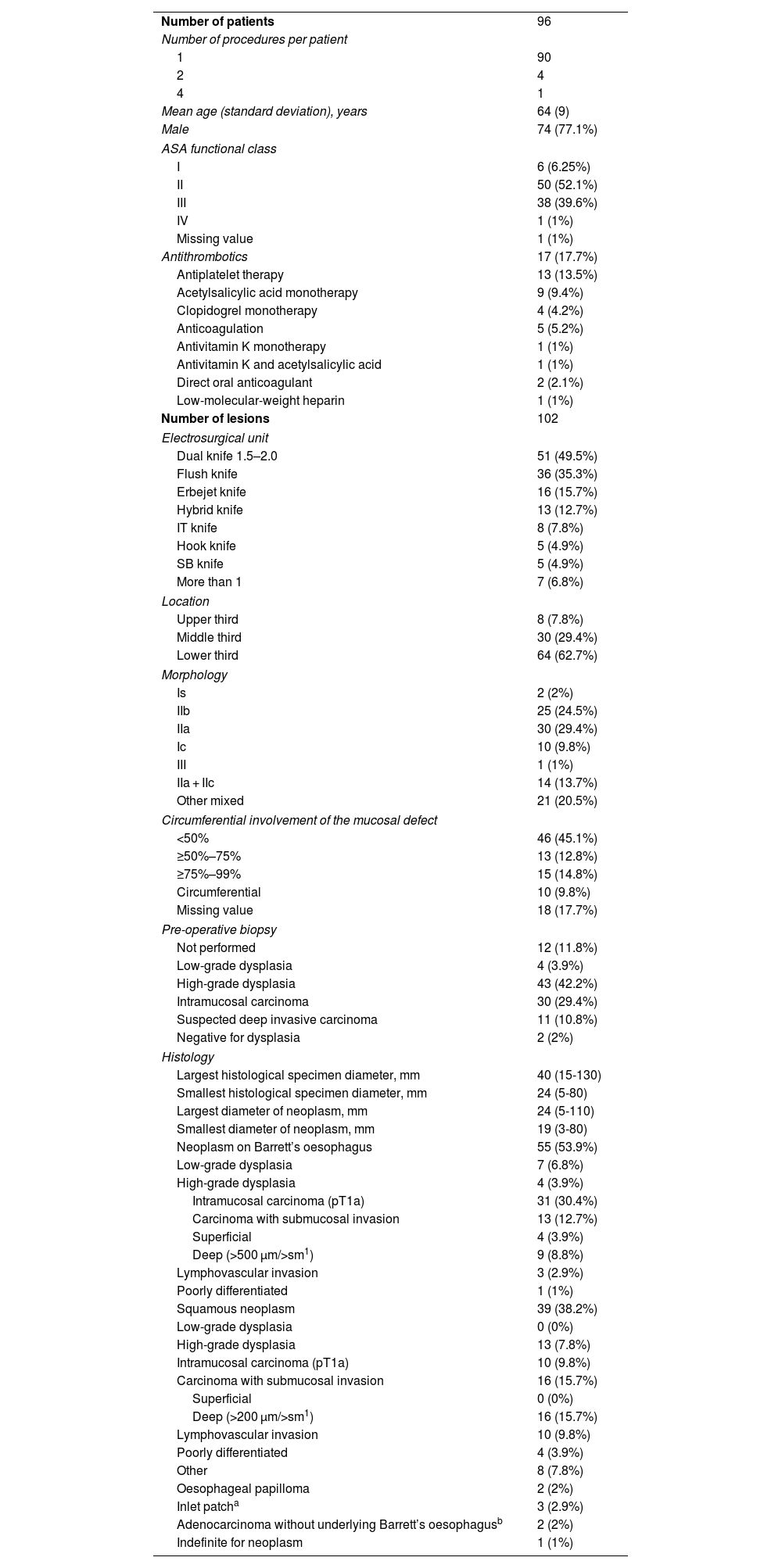
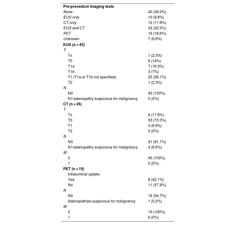
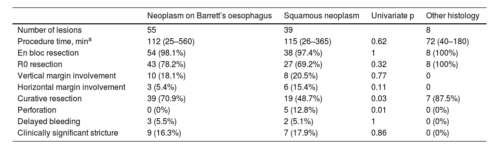
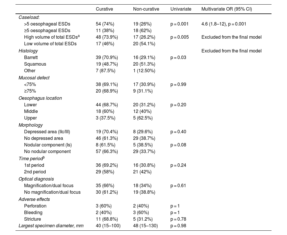
Results

A total of 102 ESD were performed on 96 patients. The technical success rate was 100% and the percentage of en-bloc resection was 98%. The percentage of R0 and curative resection was 77.5% (n = 79; 95% CI: 68%–84%) and 63.7% (n = 65; 95% CI: 54%–72%), respectively. The most frequent histology was Barrett-related neoplasia (n = 55 [53.9%]). The main reason for non-curative resection was deep submucosal invasion (n = 25). The centers with a lower volume of ESD obtained worse results in terms of curative resection. The rate of perforation, delayed bleeding and post-procedural stenosis were 5%, 5% and 15.7%, respectively. No patient died or required surgery due to an adverse effect. After a median follow-up of 14 months, 20 patients (20.8%) underwent surgery and/or chemoradiotherapy, and 9 patients died (mortality 9.4%).

Conclusions

In Spain, esophageal ESD is curative in approximately two out of three patients, with an acceptable risk of adverse events.

Keywords:
Esophageal neoplasms
Endoscopy
Endoscopic submucosal dissection
Barrett esophagus
Adverse events
Resumen
Introducción y objetivos

Los resultados de la disección submucosa endoscópica (DSE) en el esófago no han sido evaluados en nuestro país. Nuestro objetivo principal fue analizar la efectividad y la seguridad de la técnica.

Material y métodos

Análisis del registro nacional prospectivo de DSE. Se incluyeron todas las lesiones superficiales esofágicas extirpadas mediante DSE en 17 hospitales (20 endoscopistas) entre enero de 2016 y diciembre de 2021. Se excluyeron las lesiones subepiteliales. La variable principal fue el porcentaje de resección curativa. Se realizó un análisis de regresión logística para conocer los predictores de resección no curativa y un análisis de supervivencia.

Resultados

Se realizaron un total de 102 DSE en 96 pacientes. El éxito técnico fue del 100% y el porcentaje de resección en bloque, del 98%. El porcentaje de resección R0 y curativa fue del 77,5% (n = 79; IC 95%: 68%–84%) y del 63,7% (n = 65; IC 95%: 54%–72%), respectivamente. La histología más frecuente fue la neoplasia sobre esófago de Barrett (n = 55 [53,9%]). El principal motivo de resección no curativa fue la invasión submucosa profunda (n = 25). Los centros con menor volumen de casos obtuvieron cifras inferiores de resección curativa. El porcentaje de perforación, sangrado diferido y estenosis posprocedimiento fue del 5%, del 5% y del 15,7%, respectivamente. Ningún paciente falleció ni requirió cirugía por un efecto adverso. Tras una mediana de seguimiento de 14 meses, 20 pacientes (20,8%) recibieron cirugía y/o quimio-radioterapia, y 9 fallecieron (mortalidad del 9,4%).

Conclusiones

En nuestro medio, la DSE esofágica es curativa en aproximadamente dos de cada tres pacientes, con un riesgo aceptable de efectos adversos.

Palabras clave:
Cáncer de esófago
Endoscopia
Disección endoscópica submucosa
Esófago de Barrett
Eventos adversos
Full Text
Introduction

Oesophageal cancer is the seventh leading cause of cancer deaths worldwide.1 In Spain, it is estimated that the number of annual cases will increase by approximately 20% over the next 20 years.1 The classic treatment for this disease has been surgery and chemoradiotherapy. In the last decade, a large number of Asian studies have confirmed that endoscopic submucosal dissection (ESD) offers favourable results in well-selected patients in both Barrett’s oesophagus (BO) and squamous lesions.2–4 In fact, recent clinical practice guidelines recommend ESD as the first-line treatment for oesophageal neoplasms affecting the mucosa or superficial submucosa, with a low risk of regional lymphatic involvement.1,5 Compared to conventional endoscopic mucosal resection (EMR), ESD achieves greater success in terms of en bloc resection and free margins, resulting in a more accurate histological and prognostic assessment and a lower risk of recurrence.5 Additionally, case series have been reported suggesting that ESD may be indicated in less common diseases, such as adenocarcinoma arising in an inlet patch or oesophageal papillomas.6–8

Implementation of the technique in the West has been slower, probably due to a lower incidence of oesophageal cancer, lower detection of early lesions and the technical complexity of ESD.9 In addition, the risk of adverse effects is higher than with EMR, especially at the beginning of the learning curve.10 Western published results are mixed, and generally below Asian standards and those set by the European Society of Gastrointestinal Endoscopy (ESGE).11–13 In Spain, gastric ESD has been shown to yield favourable results,14 but no data have been published on the results of oesophageal ESD.

The main objective of our study was to describe the effectiveness and safety of oesophageal ESD in Spain. Our secondary objective was to analyse factors associated with non-curative resection and patient survival.

Material and methodsStudy design and population

This study was conducted in accordance with the STROBE recommendations for observational studies.15 All superficial oesophageal lesions included in the national ESD registry between January 2016 and December 2021 were analysed. Subepithelial lesions were excluded. The procedures were performed with high-definition endoscopes by 20 endoscopists in 17 different hospitals. All endoscopists were dedicated to EMR and ESD, had performed a minimum of 20 ESD in animal models and had previous experience in gastric and/or rectal ESD. The technique and dissection material used were at the discretion of each endoscopist. Furthermore, the need for complementary treatment (surgery, radiotherapy or chemotherapy) was agreed in the multidisciplinary committees of the participating institutions. The national ESD registry has received ethics committee approval from all participating hospitals, and all patients signed an informed consent for inclusion.

Data collection

The national registry prospectively collected ESDs performed in 35 Spanish hospitals, 17 of which had performed at least one case of oesophageal ESD during the study period. The information was collected on the REDCap “i + 12 Research Institute” platform of the Hospital Universitario 12 de Octubre in Madrid using a standardised case report form that included the following variables: a) demographics; b) comorbidity and antithrombotic medication; c) type of sedation; d) lesion characteristics (location, percentage of circumferential involvement, morphology according to Paris classification, optical diagnosis); e) procedure variables (technique, operator, time, manoeuvrability, en bloc resection and consumables used); f) histology (type of lesion, size, degree of differentiation, lymphovascular involvement, vertical and horizontal margins and degree of invasion); g) adverse effects; and h) follow-up (endoscopic, imaging tests and need for complementary treatments). Following peer review of the manuscript, data related to pre-procedural staging (endoscopic ultrasound [EUS], computed tomography [CT] and positron emission tomography [PET]) were retrospectively retrieved. The results of these tests are not collected prospectively in the registry and were not performed systematically in all patients, as recommended by the latest ESGE guideline.5

For this study, an additional review of the data was conducted between January and April 2022. This review consisted of confirmation of the primary endpoint and outliers, an update of the follow-up and a review of all missing values.

Study variables and definitions

The primary endpoint of the study was the percentage of curative resection, defined as an R0 resection and absence of histological criteria for poor prognosis according to the latest ESGE clinical practice guideline,5 Histological criteria for poor prognosis were considered to be lymphovascular or perineural invasion, vertical or horizontal margin involvement (<1 mm), a poor degree of differentiation and deep submucosal invasion (>500 μm in BO or >200 µm in squamous lesions or invasion sm2).5 R0 resection was defined as resection with vertical and horizontal margins free (>1 mm) of dysplasia. In patients with BO, an R0 resection was defined as when the vertical and horizontal margins were free of the lesion with more advanced histology.12 A non-curative resection with a high risk of lymph node metastasis (risk >3%) was defined according to the ESGE guideline,5 i.e. as a resection with one or more histological criteria of poor prognosis with the exception of an affected horizontal margin. A non-curative resection with a high risk of local recurrence (risk >10%) was considered to be a resection with an affected horizontal margin without other poor prognostic criteria.5 All specimens were fixed with pins and immersed in formalin after ESD for histological analysis. Histology was evaluated at each centre by an experienced pathologist specialising in gastrointestinal lesions.

Technical success was defined as endoscopic removal of the lesion of interest. Intraprocedural bleeding was defined as bleeding in oozing or pulsatile form that required specific endoscopic treatment (clips, coagulation clamp, sclerotherapy, topical agent, etc.), interventional radiology or surgery. Thirty-day delayed bleeding was defined as bleeding that met any of the following criteria: a) haematemesis or melaena after completion of the procedure; b) development of anaemia >2 g/dl haemoglobin; or c) endoscopic, radiological or surgical procedure for suspected bleeding after completion of ESD.14 Intraprocedural perforation was defined as deep muscle damage with visualisation of the longitudinal muscle, without necessarily requiring visualisation of a transmural hole. Delayed perforation was diagnosed on the basis of the clinical signs and symptoms and compatible imaging tests.14 Stricture was considered clinically significant as long as the patient reported dysphagia and required at least one endoscopic dilation.

Statistical analysis

Descriptive analysis of the quantitative variables was performed using the mean and standard deviation, or the median and range for variables with asymmetrical distribution. The qualitative variables are presented as absolute and relative frequencies. The 95% confidence intervals (CI) of the proportions were calculated using the Wilson method. Univariate analysis was performed using the chi-square (χ2) or Fisher’s method for qualitative variables, and the ANOVA or Kruskal–Wallis method for quantitative variables. Predictors of non-curative resection were analysed by multivariate binary logistic regression analysis using the all possible equations method.16 In order to minimise type I error and model overfitting, only variables that could have a reasonable clinical or pathophysiological association with the event of interest were selected as candidate variables for the final model. The decision to set a cut-off point of five cases of oesophageal ESD to categorise high-caseload and low-caseload centres was made in order to compare the first quartile with the remaining three quartiles. Disease-free survival and overall survival was estimated using the Kaplan–Meier method. Patients were right-censored at the time of the last available medical visit.

No a priori sample size estimation was made because the main objective of the study is descriptive. Missing values are presented in the tables and no imputation techniques were used. All analyses were two-tailed. A p-value of <0.05 was considered statistically significant. Statistical analysis was performed using Stata version 14.2 (StataCorp, College Station, TX, USA). All authors had access to the final study database.

Results

During the study period, 108 oesophageal ESDs were performed out of a total of 2222 ESDs (4.9%) in the 17 participating centres. Six subepithelial lesions were excluded, so the final sample consisted of 102 procedures in 96 patients (Fig. 1 and Table 1). The mean age was 64 years and the majority (77.1%) were male. In total, 41% of patients had significant comorbidity (American Society of Anesthesiologists [ASA] functional class ≥ III), and 17% were receiving antithrombotic medication.

Figure 1.

Flowchart of the study.

Table 1.

Characteristics of the population.

Number of patients  96 
Number of procedures per patient
90 
Mean age (standard deviation), years  64 (9) 
Male  74 (77.1%) 
ASA functional class
6 (6.25%) 
II  50 (52.1%) 
III  38 (39.6%) 
IV  1 (1%) 
Missing value  1 (1%) 
Antithrombotics  17 (17.7%) 
Antiplatelet therapy  13 (13.5%) 
Acetylsalicylic acid monotherapy  9 (9.4%) 
Clopidogrel monotherapy  4 (4.2%) 
Anticoagulation  5 (5.2%) 
Antivitamin K monotherapy  1 (1%) 
Antivitamin K and acetylsalicylic acid  1 (1%) 
Direct oral anticoagulant  2 (2.1%) 
Low-molecular-weight heparin  1 (1%) 
Number of lesions  102 
Electrosurgical unit
Dual knife 1.5–2.0  51 (49.5%) 
Flush knife  36 (35.3%) 
Erbejet knife  16 (15.7%) 
Hybrid knife  13 (12.7%) 
IT knife  8 (7.8%) 
Hook knife  5 (4.9%) 
SB knife  5 (4.9%) 
More than 1  7 (6.8%) 
Location
Upper third  8 (7.8%) 
Middle third  30 (29.4%) 
Lower third  64 (62.7%) 
Morphology
Is  2 (2%) 
IIb  25 (24.5%) 
IIa  30 (29.4%) 
Ic  10 (9.8%) 
III  1 (1%) 
IIa + IIc  14 (13.7%) 
Other mixed  21 (20.5%) 
Circumferential involvement of the mucosal defect
<50%  46 (45.1%) 
≥50%–75%  13 (12.8%) 
≥75%–99%  15 (14.8%) 
Circumferential  10 (9.8%) 
Missing value  18 (17.7%) 
Pre-operative biopsy
Not performed  12 (11.8%) 
Low-grade dysplasia  4 (3.9%) 
High-grade dysplasia  43 (42.2%) 
Intramucosal carcinoma  30 (29.4%) 
Suspected deep invasive carcinoma  11 (10.8%) 
Negative for dysplasia  2 (2%) 
Histology
Largest histological specimen diameter, mm  40 (15-130) 
Smallest histological specimen diameter, mm  24 (5-80) 
Largest diameter of neoplasm, mm  24 (5-110) 
Smallest diameter of neoplasm, mm  19 (3-80) 
Neoplasm on Barrett’s oesophagus  55 (53.9%) 
Low-grade dysplasia  7 (6.8%) 
High-grade dysplasia  4 (3.9%) 
Intramucosal carcinoma (pT1a)  31 (30.4%) 
Carcinoma with submucosal invasion  13 (12.7%) 
Superficial  4 (3.9%) 
Deep (>500 μm/>sm19 (8.8%) 
Lymphovascular invasion  3 (2.9%) 
Poorly differentiated  1 (1%) 
Squamous neoplasm  39 (38.2%) 
Low-grade dysplasia  0 (0%) 
High-grade dysplasia  13 (7.8%) 
Intramucosal carcinoma (pT1a)  10 (9.8%) 
Carcinoma with submucosal invasion  16 (15.7%) 
Superficial  0 (0%) 
Deep (>200 μm/>sm116 (15.7%) 
Lymphovascular invasion  10 (9.8%) 
Poorly differentiated  4 (3.9%) 
Other  8 (7.8%) 
Oesophageal papilloma  2 (2%) 
Inlet patcha  3 (2.9%) 
Adenocarcinoma without underlying Barrett’s oesophagusb  2 (2%) 
Indefinite for neoplasm  1 (1%) 

ASA: American Society of Anesthesiologists.

Quantitative variables were expressed in terms of median and range.

a

Includes two patients with adenocarcinoma and one patient with pre-intervention dysplasia not confirmed on histology.

b

One of the patients with adenocarcinoma without Barrett’s oesophagus had deep submucosal invasion (1800 μm).

Procedures

The distribution of the number of cases per hospital was asymmetrical (Fig. 2). Five centres with more than five cases accounted for 72% (n = 73) of the procedures. An average of one ESD procedure was performed per centre per year. The median procedure time was 111 min (range: 25–560). Most procedures were performed using CO2 (n = 99 [97.1%]) and with orotracheal intubation (n = 96 [94.1%]).

Figure 2.

Number of cases per centre.

The technical success rate was 100% and the en bloc resection rate was 98% (n = 100; 95% CI: 93.1%–99.4%). Magnification was used in 22 lesions (21.5%) and dual focus technology in 47 lesions (46.1%) to assess the resectability of the lesion. The most commonly used scalpels were the Dual knife (Olympus Corp., Tokyo, Japan) and the Flush knife (Fujifilm Corp., Tokyo, Japan) (Table 1). Some form of traction was used in 34 lesions (33.3%), the tunnelling technique in 27 (26.4%), and both tunnelling and traction in seven (6.9%).

Pre-procedural staging

Pre-ESD biopsies were obtained in 88.2% (n = 90) of the procedures (Table 1). The degree of dysplasia between pre-ESD biopsy and final histology was concordant in 38.9% of lesions (n = 35), underestimated in 37.8% (n = 34) and overestimated in 23.3% (n = 21). In 40 lesions (39.2%), no imaging tests were performed prior to the procedure. The number of imaging tests performed (EUS/CT/PET) and their results are detailed in Table 2.

Table 2.

Staging imaging tests.

Pre-procedure imaging tests
None  40 (39.2%) 
EUS only  10 (9.8%) 
CT only  12 (11.8%) 
EUS and CT  33 (32.3%) 
PET  19 (18.6%) 
Unknown  7 (6.9%) 
EUS (n = 43)
T
Tx  1 (2.3%) 
T0  6 (14%) 
T1a  7 (16.3%) 
T1b  3 (7%) 
T1 (T1a or T1b not specified)  25 (58.1%) 
T2  1 (2.3%) 
N
N0  43 (100%) 
N1/adenopathy suspicious for malignancy  0 (0%) 
CT (n = 45)
T
Tx  8 (17.8%) 
T0  33 (73.3%) 
T1  4 (8.9%) 
T2  0 (0%) 
N
N0  41 (91.1%) 
N1/adenopathy suspicious for malignancy  4 (8.9%) 
M
45 (100%) 
0 (0%) 
PET (n = 19)
Intraluminal uptake
Yes  8 (42.1%) 
No  11 (57.9%) 
N
N0  18 (94.7%) 
Adenopathies suspicious for malignancy  1 (5.2%) 
M
19 (100%) 
0 (0%) 

CT: computerised axial tomography; PET: positron emission tomography.

Lesion characteristics and histology

The morphological and histological characteristics of the lesions are detailed in Table 1. The most frequent location was the distal oesophagus (62.7%). Some 53.9% (n = 55) of the dissections were performed on a BO and in 38.2% (n = 39) the histology was squamous in nature. Intramucosal and deep submucosal invasive neoplasms were the most frequent histologies.

Resection with free margins and curative resection

The percentage of R0 and curative resection was 77.5% (n = 79, 95% CI: 68%–84%) and 63.7% (n = 65, 95% CI: 54%–72%), respectively. The most frequent reason for non-curative resection (n = 37) was deep submucosal invasion (n = 25 [24%]), followed by vertical margin involvement (n = 18 [17.5%]), lymphovascular invasion (n = 13 [12.7%]), horizontal margin involvement (n = 9 [8.8%]), undifferentiated tumour (n = 5 [5.9%]) and fragmented resection (n = 2 [2%]). In 16 patients (15.7%) there were two or more coexisting factors for non-curative resection. Following the criteria of the ESGE guideline,5 31 patients had a non-curative resection with a high risk of lymph node metastasis and six with a high risk of local recurrence.

The results according to histology (BO versus squamous) are detailed in Table 3. In the univariate analysis, centres with a smaller caseload (n ≤ 5 oesophageal ESD) and squamous histology were associated with a lower percentage of curative resection. A smaller ESD caseload in any location was also associated with this variable, but was excluded from the final model due to collinearity with the number of oesophageal ESDs. In the multivariate analysis, the only factor associated with an increased risk of non-curative resection was that the ESD was performed in a hospital with fewer procedures (≤5 oesophageal ESD) (Table 4).

Table 3.

Results according to histology (squamous vs Barrett’s oesophagus).

  Neoplasm on Barrett’s oesophagus  Squamous neoplasm  Univariate p  Other histology 
Number of lesions  55  39   
Procedure time, mina  112 (25–560)  115 (26–365)  0.62  72 (40–180) 
En bloc resection  54 (98.1%)  38 (97.4%)  8 (100%) 
R0 resection  43 (78.2%)  27 (69.2%)  0.32  8 (100%) 
Vertical margin involvement  10 (18.1%)  8 (20.5%)  0.77 
Horizontal margin involvement  3 (5.4%)  6 (15.4%)  0.11 
Curative resection  39 (70.9%)  19 (48.7%)  0.03  7 (87.5%) 
Perforation  0 (0%)  5 (12.8%)  0.01  0 (0%) 
Delayed bleeding  3 (5.5%)  2 (5.1%)  0 (0%) 
Clinically significant stricture  9 (16.3%)  7 (17.9%)  0.86  0 (0%) 

The univariate analysis compares squamous neoplasms vs Barrett’s oesophagus. Lesions with other histology have been excluded from the comparison due to the low number of cases.

a

Median and range.

Table 4.

Predictors of non-curative resection.

  Curative  Non-curative  Univariate  Multivariate OR (95% CI) 
Caseload:
>5 oesophageal ESDs  54 (74%)  19 (26%)  p = 0.001  4.6 (1.8–12), p = 0.001 
≥5 oesophageal ESDs  11 (38%)  18 (62%)     
High volume of total ESDsa  48 (73.9%)  17 (26.2%)  p = 0.005  Excluded from the final model 
Low volume of total ESDs  17 (46%)  20 (54.1%)     
Histology        Excluded from the final model 
Barrett  39 (70.9%)  16 (29.1%)  p = 0.03   
Squamous  19 (48.7%)  20 (51.3%)     
Other  7 (87.5%)  1 (12.50%)     
Mucosal defect
<75%  38 (69.1%)  17 (30.9%)  p = 0.99   
≥75%  20 (68.9%)  9 (31.1%)     
Oesophagus location
Lower  44 (68.7%)  20 (31.2%)  p = 0.20   
Middle  18 (60%)  12 (40%)     
Upper  3 (37.5%)  5 (62.5%)     
Morphology
Depressed area (IIc/III)  19 (70.4%)  8 (29.6%)  p = 0.40   
No depressed area  46 (61.3%)  29 (38.7%)     
Nodular component (Is)  8 (61.5%)  5 (38.5%)  p = 0.08   
No nodular component  57 (66.3%)  29 (33.7%)     
Time periodb
1st period  36 (69.2%)  16 (30.8%)  p = 0.24   
2nd period  29 (58%)  21 (42%)     
Optical diagnosis
Magnification/dual focus  35 (66%)  18 (34%)  p = 0.61   
No magnification/dual focus  30 (61.2%)  19 (38.8%)     
Adverse effects
Perforation  3 (60%)  2 (40%)  p = 1   
Bleeding  2 (40%)  3 (60%)  p = 1   
Stricture  11 (68.8%)  5 (31.2%)  p = 0.78   
Largest specimen diameter, mm  40 (15–100)  48 (15–130)  p = 0.98   

CI: confidence interval; ESD: endoscopic submucosal dissection; OR: odds ratio.

Quantitative variables were expressed in terms of median and range. Percentages represent row percentages.

a

The 17 participating centres were divided into 4 groups according to the number of total ESD cases performed (see Fig. 2). The five centres with the highest number of procedures were considered to be large caseload centres (first quartile). All these centres performed more than 140 ESDs during the study period. This variable was not considered for the final multivariate model due to collinearity with the number of oesophageal ESDs.

b

To assess the potential impact of the learning curve at each centre, the procedures performed at each institution were divided into two time periods (50% of cases in each period).

Adverse effects

In total, 44.1% (n = 45) of patients experienced an adverse effect. Intraprocedural bleeding was the most frequent (n = 39 [38.5%]). Five patients (4.9%) had delayed bleeding after a median of seven days (range: 1–13). All intraprocedural and delayed bleeding was controlled endoscopically. Intraprocedural perforation occurred in five patients (4.9%) and no delayed perforation was observed. All perforations occurred in ESDs of squamous neoplasms (univariate p = 0.03, Table 3) and were successfully treated with haemoclips. In four of these perforations, neither the tunnelling technique nor any kind of traction had been used.

To prevent the occurrence of stricture, some form of prophylaxis was performed in 26 lesions (25.5% of procedures: n = 13 oral corticosteroids, n = 7 topical corticosteroids, n = 1 intravenous corticosteroids and n = 5 combined treatment). Prophylaxis was used more frequently in patients with a circumferential mucosal defect ≥75% (64% vs 15.2%; p < 0.001). The median oral corticosteroid starting dose was 30 mg (range 30–80), with a median treatment duration of eight weeks (range 1–12). The median dose of topical corticosteroid injected into the eschar was 80 mg (range 40–480). Dysphagia requiring a median of three dilation sessions (range: 1–13) occurred in 16 patients (15.7%). The occurrence of clinically significant stricture was more frequent in patients with a circumferential mucosal defect ≥75% (36% vs 5%; p < 0.001). The use of corticosteroids as prophylaxis did not decrease the risk of developing a stricture (75% patients with prophylaxis vs 17% without prophylaxis; p = 0.001). After adjusting for circumferential extent and specimen size, no significant differences were found (p = 0.08). Dysphagia resolved in all but three patients: two patients required stent placement and one patient underwent oncologically-indicated surgery due to non-curative resection.

The median length of hospital stay was two days (range: 0–7) for patients who did not experience an adverse event and three days (range: 1–15) for those who experienced one or more complications.

Follow-up

Median follow-up was 14 months (range: 1–66). In four patients (4.2%), no follow-up visits were recorded after discharge from hospital.

Of the 31 patients with non-curative resection and high risk of lymph node metastasis, five were treated with surgery, four with surgery and chemoradiotherapy, four with chemoradiotherapy, six with radiotherapy alone, one with chemotherapy alone, and in 11 no adjuvant treatment was administered due to the decision of the multidisciplinary committee looking after the patient. Of the nine patients who underwent surgery, two had no residual disease in the surgical specimen, four had nodal disease and three had intraluminal disease. Of the 11 patients at high risk of lymph node metastasis in whom no adjuvant treatment was given, one patient died of non-cancerous disease and two developed distant disease after a median follow-up of 11 months.

Nine deaths occurred during follow-up (overall mortality: 9/96 [9.4%]), four of them secondary to tumour disease. Overall survival and disease-free survival at two years was 89% (95% CI: 76%–95%) and 80% (95% CI: 66%–89%), respectively. Disease-free survival was longer in patients in whom curative resection was achieved (logrank p < 0.01), with no statistically significant difference in overall survival (five deaths in the curative resection group and four in the non-curative group, logrank p = 0.24). Five patients developed metastatic disease after a median of 11 months post-ESD (range 4–15).

Discussion

The ESGE guideline suggests that the en bloc resection rate should be greater than 90%, the cure rate over 75%, the risk of perforation less than 3% and the need for emergency surgery less than 1%.10 Analysis of the national and prospective ESD registry indicates that the technical success and en bloc resection figures (98%) are excellent, with an acceptable incidence of adverse effects. However, the curative resection rate (64%) is below desirable, mainly due to the suboptimal results found in ESD of squamous neoplasms (49% curative resection).11

The results of the main published series of oesophageal ESD are summarised in Appendix B, Supplementary Table 1. Interpretation of our results requires an analysis stratified by histology (squamous or BO). ESD in BO is positioned as first-line treatment for lesions with suspected submucosal invasion, i.e. those larger than 2 cm, with nodular or depressed morphology (0-Is or 0-IIc of the Paris classification) and for those with anticipated submucosal fibrosis.5 In this clinical scenario, the curative resection and safety results in our cohort are in line with those published in other Western studies.12,17 In a 2018 meta-analysis analysing the outcomes of ESD in BO, the rate of en bloc resection, R0 resection and curative resection was 92.9% (95% CI: 90.3%–95.2%), 74.5% (95% CI: 66.3%–81.9%) and 64.9% (95% CI: 55.7%–73.6%), respectively.3 Subsequent North American and European series show similar findings.12,17 In the context of squamous cell carcinoma, published data are available from France and Germany with which to compare our results. The Western study with the largest sample size is the French cohort dating from 2019.11 Berger et al.11 compared the results of EMR and ESD in 148 lesions (80 EMR vs 68 ESD). ESD was superior in terms of curative resection (74% vs 21%; p < 0.001) and five-year disease-free survival (95% vs 73%; p = 0.01), success rates higher than those found in our study. In contrast, the results of the recently published German registry are similar to ours in terms of curability (n = 54 squamous lesions, 46.2% curative resection).18

The design of our study does not allow us to establish the causes of the lower success rate in squamous lesions, but several hypotheses can be postulated. The high rate of en bloc resection (98%) suggests that the main reason is probably not a technical problem but poor lesion selection. Firstly, it is important to note that the main reason for non-curative resection was deep submucosal invasion. Endoscopists’ unfamiliarity with optical diagnosis of squamous lesions and lack of training in the use of magnification to estimate the risk of submucosal invasion may have played a role. As recommended by the latest ESGE guideline,5 systematic use of the Japanese Oesophageal Society classification could improve outcomes. This classification analyses the morphology and loop arrangement of the papillary capillaries in the mucosa to estimate the risk of submucosal invasion. Although there are no Western studies, several Japanese series suggest that its sensitivity and specificity are around 85%–90% after suitable training.19 Secondly, it is important to note that approximately 40% of our cohort had significant comorbidity (ASA ≥ III). It is possible that excision by ESD was chosen despite lesions with significant risk of deep submucosal invasion, as oesophagectomy is associated with increased morbidity and mortality. The morbidity and mortality of the surgical alternative in the oesophagus is not comparable to surgery in other anatomical locations, such as right hemicolectomy or antrectomy. Thirdly, our secondary analysis suggests that a smaller caseload has a negative impact on technique outcome. A lower number of total ESDs or oesophageal ESDs was statistically significantly associated with a higher risk of non-curative resection. This is a constant in advanced endoscopic procedures that have a long learning curve.10,20,21 These findings, together with the small caseload per year (average of one case per centre per year), suggest the need to create referral centres and underline the importance of maintaining a significant volume of cases to safeguard competence, especially in a country with a low incidence of oesophageal cancer such as Spain.

Concerns about the increased risk of adverse effects and the lack of standardisation of the technique are some of the barriers limiting the widespread implementation of ESD in our setting. Although the figures for perforation (5%) and bleeding (5%) are slightly higher than those found in other non-Asian series,11,12,17,18 we believe they are within an acceptable range (Appendix B, Supplementary Table 1). It should be noted that no patient died or required surgery due to an adverse effect, as recommended by the ESGE.10 Approximately one third of our series used some form of traction or submucosal tunnelling endoscopic dissection. A wider implementation of these technical variants could help to reduce the incidence of complications and procedure time, as reported in several observational studies and clinical trials.22,23

Stricture is another major concern in oesophageal ESD, occurring in 2%–15% of cases.3,13 Postulated risk factors are the location in the upper oesophagus, the longitudinal and circumferential extent of the mucosal defect, the flat and depressed morphology and the degree of deep invasion.24 The significant prevalence in our cohort of lesions with a mucosal defect greater than 75% and the failure to use prophylaxis in a significant percentage of these lesions (36%) may explain why the incidence of stricture in our study is at the higher end of the published range. The use of corticosteroids (oral and/or topical), metal stents and other topical substances may prevent the risk of developing stricture, although the results are disputed.25 There is no high-quality evidence to support a specific treatment algorithm, which explains the heterogeneity of the prophylactic guidelines used in our series. The Japanese clinical practice guideline makes a weak recommendation in favour of using topical corticosteroids in lesions involving more than 75%,19 while the European guideline does not specify an algorithm.5

The strengths of our study include its prospective nature, the fact that it reflects the experience of the technique across Spain, and a sample size similar to other European series. However, it also has limitations. Firstly, the finding of the logistic regression analysis on the impact of caseload should be interpreted as a hypothesis. The number of cases collected does not allow for a multilevel analysis adjusting for individual endoscopists and hospitals. Secondly, patient follow-up is still limited and is continuously being updated: five-year results are needed to assess the technique more reliably in oncological terms. Thirdly, eight heterogeneous lesions (e.g., squamous papillomas, lesion arising in an inlet patch, etc.) that are often excluded in other cohorts have been included. The rationale for their inclusion lies in the importance of generating knowledge about these conditions that are often reported as clinical cases6,7 and for which ESD may represent a therapeutic alternative. Finally, a centralised assessment of histology was not performed.

In conclusion, initial experience with oesophageal ESD in Spain indicates that the technique is curative in approximately two out of three patients, with an acceptable safety profile. For lesions on BO, the figures are in line with Western series, but there is significant room for improvement for squamous lesions. The significant number of lesions with margin involvement and/or deep submucosal invasion suggests that better lesion assessment would contribute to improved outcomes.

Funding

No specific funding was received for this study. The Spanish registry of endoscopic submucosal dissection has received a grant from the Sociedad Española de Endoscopia Digestiva [Spanish Society of Gastrointestinal Endoscopy].

Conflicts of interest

Enrique Rodríguez de Santiago declares having received fees from Olympus (training and advisory activities), Norgine (registration to courses and conferences) and Casen Recordati (registration to courses and conferences).

José Carlos Marín-Gabriel declares having received fees from Olympus, Fujifilm, Norgine and Casen Recordati (training activities).

Alberto Herreros de Tejada declares having received fees from Olympus, Fujifilm, Norgine, Creo Medical, Boston Scientific, Izasa, 3D-Matrix, Ambu and Casen Recordati (training and/or consultancy activities).

Hugo Uchima declares having received fees from Izasa, ERBE-Spain and Ambu.

The other authors declare that they have no conflicts of interest related to this publication.

Appendix A
Supplementary data

The following is Supplementary data to this article:

Icono mmc1.doc

References
[1]
A. Fernández-Montes, J. Alcaide, M. Alsina, A.B. Custodio, L.F. Franco, J. Gallego Plazas, et al.
SEOM-GEMCAD-TTD clinical guideline for the diagnosis and treatment of esophageal cancer (2021).
Clin Transl Oncol, 24 (2022), pp. 658-669
[2]
H. Noguchi, Y. Naomoto, H. Kondo, M. Haisa, T. Yamatsuji, K. Shigemitsu, et al.
Evaluation of endoscopic mucosal resection for superficial esophageal carcinoma.
Surg Laparosc Endosc Percutan Tech, 10 (2000), pp. 343-350
[3]
D. Yang, F. Zou, S. Xiong, J.J. Forde, Y. Wang, P.V. Draganov.
Endoscopic submucosal dissection for early Barrett’s neoplasia: a meta-analysis.
Gastrointest Endosc, 87 (2018), pp. 1383-1393
[4]
Z. Liu, R. Zhao.
Endoscopic submucosal dissection vs. surgery for superficial esophageal squamous cancer: a systematic review and meta-analysis.
Front Oncol, 12 (2022), pp. 816832
[5]
P. Pimentel-Nunes, D. Libânio, B.A.J. Bastiaansen, P. Bhandari, R. Bisschops, M.J. Bourke, et al.
Endoscopic submucosal dissection for superficial gastrointestinal lesions: European Society of Gastrointestinal Endoscopy (ESGE) guideline — update 2022.
Endoscopy, 54 (2022), pp. 591-622
[6]
R. Rusu, S. Ishaq, T. Wong, J.M. Dunn.
Cervical inlet patch: new insights into diagnosis and endoscopic therapy.
Frontline Gastroenterol, 9 (2018), pp. 214-220
[7]
J.Y. Cho, D.Y. Cheung, T.J. Kim, J.K. Kim.
A case of esophageal squamous cell carcinoma in situ arising from esophageal squamous papilloma.
Clin Endosc, 52 (2019), pp. 72-75
[8]
A. Probst, T. Schaller, H. Messmann.
Adenocarcinoma arising from ectopic gastric mucosa in an esophageal inlet patch: treatment by endoscopic submucosal dissection.
Endoscopy, 47 (2015), pp. E337-E338
[9]
M.X. Ma, M.J. Bourke.
Endoscopic submucosal dissection in the West: current status and future directions.
Dig Endosc, 30 (2018), pp. 310-320
[10]
P. Pimentel-Nunes, M. Pioche, E. Albéniz, F. Berr, P. Deprez, A. Ebigbo, et al.
Curriculum for endoscopic submucosal dissection training in Europe: European Society of Gastrointestinal Endoscopy (ESGE) position statement.
Endoscopy, 51 (2019), pp. 980-992
[11]
A. Berger, G. Rahmi, G. Perrod, M. Pioche, J.-M. Canard, E. Cesbron-Métivier, et al.
Long-term follow-up after endoscopic resection for superficial esophageal squamous cell carcinoma: a multicenter Western study.
Endoscopy, 51 (2019), pp. 298-306
[12]
D. Yang, W. King, H. Aihara, M.S. Karasik, S. Ngamruengphong, A.A. Aadam, et al.
Effect of endoscopic submucosal dissection on histologic diagnosis in Barrett’s esophagus visible neoplasia.
Gastrointest Endosc, 95 (2022), pp. 626-633
[13]
D.C. Daoud, N. Suter, M. Durand, M. Bouin, B. Faulques, D. von Renteln.
Comparing outcomes for endoscopic submucosal dissection between Eastern and Western countries: a systematic review and meta-analysis.
World J Gastroenterol, 24 (2018), pp. 2518-2536
[14]
G. Fernández-Esparrach, J.-C. Marín-Gabriel, A.H. de Tejada, E. Albéniz, O. Nogales, A.J. del Pozo-García, et al.
Implementation of endoscopic submucosal dissection in a country with a low incidence of gastric cancer: results from a prospective national registry.
United European Gastroenterol J, 9 (2021), pp. 718-726
[15]
E. von Elm, D.G. Altman, M. Egger, S.J. Pocock, P.C. Gøtzsche, J.P. Vandenbroucke, et al.
The Strengthening the Reporting of Observational Studies in Epidemiology (STROBE) statement: guidelines for reporting observational studies.
J Clin Epidemiol, 61 (2008), pp. 344-349
[16]
J.M. Doménech, J.B. Navarro.
Find the best subset for linear, logistic and cox regression: user-written command confound for Stata [computer program].
Universitat Autònoma de Barcelona, (2020),
[17]
S.N. van Munster, E.P.D. Verheij, E.A. Nieuwenhuis, J.G.J.A. Offerhaus, S.L. Meijer, L.A.A. Brosens, et al.
Extending treatment criteria for Barrett’s neoplasia: results of a nationwide cohort of 138 endoscopic submucosal dissection procedures.
Endoscopy, 54 (2022), pp. 531-541
[18]
C. Fleischmann, A. Probst, A. Ebigbo, S. Faiss, B. Schumacher, H.-P. Allgaier, et al.
Endoscopic submucosal dissection in Europe: results of 1000 neoplastic lesions from the german endoscopic submucosal dissection registry.
Gastroenterology, 161 (2021), pp. 1168-1178
[19]
R. Ishihara, M. Arima, T. Iizuka, T. Oyama, C. Katada, M. Kato, et al.
Endoscopic submucosal dissection/endoscopic mucosal resection guidelines for esophageal cancer.
Dig Endosc, 32 (2020), pp. 452-493
[20]
S.W. Van der Merwe, R.L.J. van Wanrooij, M. Bronswijk, S. Everett, S. Lakhtakia, M. Rimbas, et al.
Therapeutic endoscopic ultrasound: European Society of Gastrointestinal Endoscopy (ESGE) guideline.
Endoscopy, 54 (2022), pp. 185-205
[21]
G. Johnson, G. Webster, I. Boškoski, S. Campos, S.K. Gölder, C. Schlag, et al.
Curriculum for ERCP and endoscopic ultrasound training in Europe: European Society of Gastrointestinal Endoscopy (ESGE) position statement.
Endoscopy, 53 (2021), pp. 1071-1087
[22]
C. Lopimpisuth, M. Simons, V.S. Akshintala, K. Prasongdee, J. Nanavati, S. Ngamruengphong.
Traction-assisted endoscopic submucosal dissection reduces procedure time and risk of serious adverse events: a systematic review and meta-analysis.
Surg Endosc, 36 (2022), pp. 1775-1788
[23]
X. Fan, Q. Wu, R. Li, W. Chen, H. Xie, X. Zhao, et al.
Clinical benefit of tunnel endoscopic submucosal dissection for esophageal squamous cancer: a multicenter, randomized controlled trial.
Gastrointest Endosc, 96 (2022), pp. 436-444
[24]
N. Lin, J. Lin, J. Gong.
Risk factors of postoperative stricture after endoscopic submucosal dissection for superficial esophageal neoplasms: a meta-analysis.
Medicine (Baltimore), 100 (2021), pp. e28396
[25]
M. Yu, Y. Tan, D. Liu.
Strategies to prevent stricture after esophageal endoscopic submucosal dissection.
Ann Transl Med, 7 (2019), pp. 271

The three authors share first authorship.

Copyright © 2023. Elsevier España, S.L.U.. All rights reserved
Article options
Tools
Supplemental materials